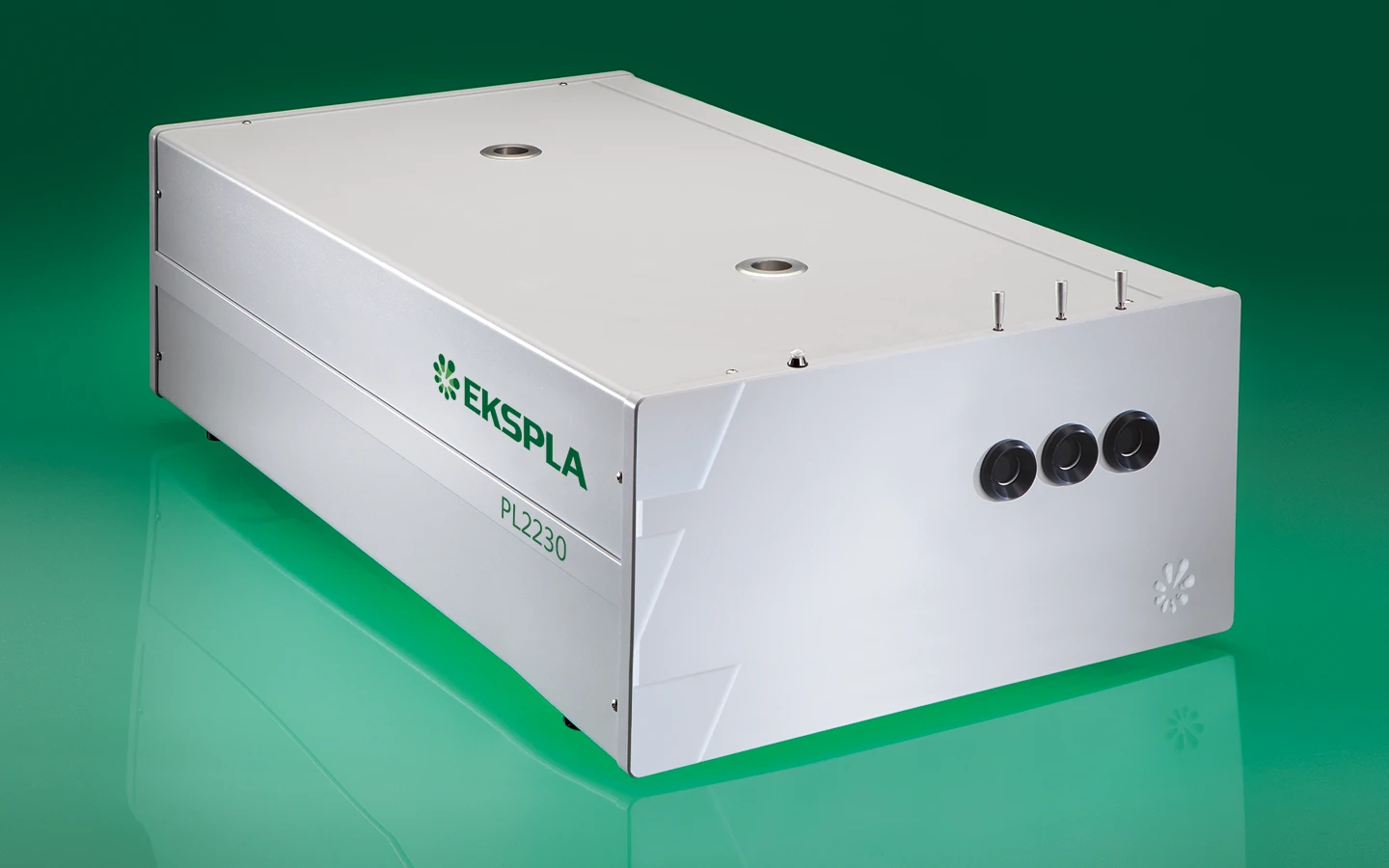
Diode Pumped High Energy Picosecond Nd:YAG Lasers PL2230
PL2230 series
Diode Pumped High Energy Picosecond Nd:YAG Lasers
PL2230 series diode-pumped, air-cooled, mode-locked picosecond Nd:YAG lasers provide high up to 40 mJ energy picosecond pulses at a 100 Hz (up to 70 mJ @ 50 Hz) pulse repetition rate. Precise pulse energy control, excellent short-term and long-term stabilit makes PL2230 series picosecond lasers an excellent choice for many demanding scientific applications.
| Model | PL2230-100 | PL2230A-100 | PL2231-50 | PL2231A-50 |
|---|---|---|---|---|
| Main specifications 1) | ||||
| Pulse energy 2) | ||||
| at 1064 nm | 3 mJ | 6 mJ | 30 mJ | 40 mJ |
| at 532 nm 3) | 1.3 mJ | 3 mJ | 13 mJ | 18 mJ |
| at 355 nm 4) | 0.9 mJ | 2 mJ | 9 mJ | 13 mJ |
| at 266 nm 5) | 0.3 mJ | 0.6 mJ | 3 mJ | 5 mJ |
| at 213 nm 6) | inquire | inquire | inquire | inquire |
| Pulse energy stability (StdDev) 7) | ||||
| at 1064 nm | < 0.2 % | < 0.6 % | < 0.5 % | < 0.5 % |
| at 532 nm | < 0.4 % | < 0.4 % | < 0.8 % | < 0.8 % |
| at 355 nm | < 0.5 % | < 0.5 % | < 1.1 % | < 1.1 % |
| at 266 nm | < 0.5 % | < 0.5 % | < 1.2 % | < 1.2 % |
| at 213 nm | < 1.5 % | < 1.5 % | < 1.5 % | < 1.5 % |
| Pulse duration (FWHM) 8) | 29 ± 5 ps | 29 ± 5 ps | 29 ± 5 ps | 29 ± 5 ps |
| Pulse duration stability 9) | ± 1 % | ± 1 % | ± 1 % | ± 1 % |
| Power drift 10) | ± 2 % | ± 2 % | ± 2 % | ± 2 % |
| Pulse repetition rate | ||||
| at 1064, 532, 355 nm | 0 – 100 Hz | 100 Hz | 50 Hz | 50 Hz |
| at 266, 213 nm | 100 Hz | 100 Hz | 10 Hz | 10 Hz |
| Polarization 11) | vertical, > 99 % | vertical, > 99 % | vertical, > 99 % | vertical, > 99 % |
| Pre-pulse contrast 12) | > 200 : 1 | > 200 : 1 | > 200 : 1 | > 200 : 1 |
| Beam profile 13) | close to Gaussian | close to Gaussian | close to Gaussian | close to Gaussian |
| Beam divergence 14) | < 1.5 mrad | < 0.7 mrad | < 0.7 mrad | < 0.7 mrad |
| Beam propagation ratio M2 | < 1.3 | < 1.3 | < 2.5 | < 2.5 |
| Beam pointing stability (RMS) 15) | ≤ 10 μrad | ≤ 20 μrad | ≤ 20 μrad | ≤ 20 μrad |
| Typical beam diameter 16) | ~ 2 mm | ~ 2.5 mm | ~ 6 mm | ~ 7 mm |
| Optical pulse jitter | ||||
| Internal triggering regime 17) | <50 ps | <50 ps | <50 ps | <50 ps |
| External triggering regime 18) | ~3 ns | ~3 ns | ~3 ns | ~3 ns |
| TRIG1 OUT pulse delay 19) | -500 … 50 ns | -500 … 50 ns | -500 … 50 ns | -500 … 50 ns |
| Typical warm-up time | 5 min | 10 min | 15 min | 15 min |
| Physical characteristics | ||||
| Laser unit size (W × L × H) | 508×1030×244 ±3 mm | 508×1030×244 ±3 mm | 508×1030×244 ±3 mm | 508×1030×244 ±3 mm |
| Electrical cabinet size (W × L × H) | 12 V DC power adapter, 85×170×41 ± 3 mm | 471×391×147 ± 3 mm | 471×391×147 ± 3 mm | 471×391×147 ± 3 mm |
| Umbilical length | 2.5 m | 2.5 m | 2.5 m | 2.5 m |
| Operating requirements | ||||
| Cooling 20) | not required, air cooled | not required, air cooled | stand-alone chiller | stand-alone chiller |
| Room temperature | 22 ± 2 °C | 22 ± 2 °C | 22 ± 2 °C | 22 ± 2 °C |
| Relative humidity | 20 – 80 % (non-condensing) | 20 – 80 % (non-condensing) | 20 – 80 % (non-condensing) | 20 – 80 % (non-condensing) |
| Power requirements | 110 – 240 V AC, 50/60 Hz | 110 – 240 V AC, 5 A, single phase 50/60 Hz | 110 – 240 V AC, 5 A, single phase 50/60 Hz | 110 – 240 V AC, 5 A, single phase 50/60 Hz |
| Power consumption | < 0.15 kVA | < 1.0 kVA | < 1.0 kVA | < 1.0 kVA |
| Model | PL2230-100 | PL2230A-100 | PL2231-50 | PL2231A-50 |
|---|
- Due to continuous improvement, all specifications are subject to change without notice. Parameters marked typical are not specifications. They are indications of typical performance and will vary with each unit we manufacture. Unless stated otherwise, all specifications are measured at 1064 nm and for basic system without options.
- Outputs are not simultaneous.
- For PL2230 series laser with –SH, -SH/TH, -SH/FH or -SH/TH/FH option or –SH/TH/FH/FiH module.
- For PL2230 series laser with –TH, -SH/TH or -SH/TH/FH option or –SH/TH/FH/FiH module.
- For PL2230 series laser with -SH/FH or -SH/TH/FH option or –SH/TH/FH/FiH module.
- For PL2230 series laser with –SH/TH/FH/FiH module.
- Averaged from pulses, emitted during 30 sec time interval.
- FWHM. Inquire for optional pulse durations in 20 – 90 ps range. Pulse energy specifications may differ from indicated here.
- Measured over 1 hour period when ambient temperature variation is less than ±1 °C.
- Measured over 8 hours period after 20 min warm-up when ambient temperature variation is less than ± 2 °C.
- At 1064 nm.
- Peak-to-peak with respect to residual pulses.
- In near and far fields. Near field Gaussian fit is >80%.
- Average of X- and Y-plane full angle divergence values measured at the 1/e2 level at 1064 nm.
- Beam pointing stability is evaluated from fluctuations of beam centroid position in the far field.
- Beam diameter is measured at 1064 nm at the 1/e2 level.
- StdDev. With respect to TRIG1 OUT pulse. <10 ps jitter is provided optionally with PRETRIG feature.
- StdDev. With respect to SYNC IN pulse.
- TRIG1 OUT lead or delay can be adjusted with 0.25 ns steps in specified range.
- Air cooled. Adequate room air conditioning should be provided.
Note: Laser must be connected to the mains electricity all the time. If there will be no mains electricity for longer that 1 hour then laser (system) needs warm up for a few hours before switching on.

Ordering information of PL2230 lasers.
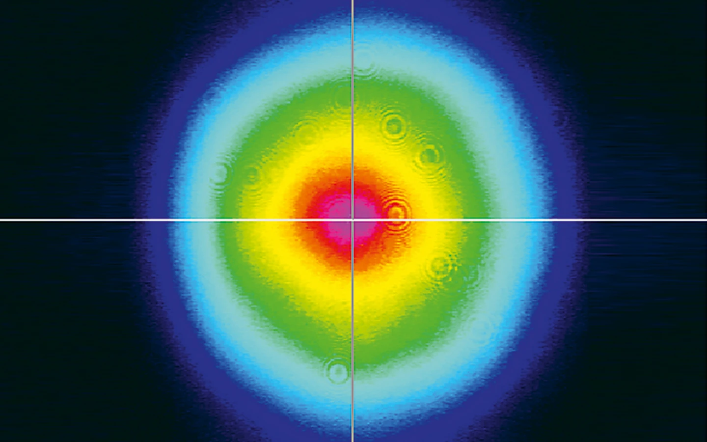
Typical near field output beam profile of PL2230 model laser.

Dimensions of PL2230 series laser head.
Ultrafast transient absorption spectra and kinetics of human blue cone visual pigment at room temperature
A. Krishnamoorthi, D. Salom, A. Wu, K. Palczewski, and P. M. Rentzepis, Proceedings of the National Academy of Sciences 121 (41), e2414037121 (2024).
DOI: 10.1073/pnas.2414037121.
Near infrared-triggered liposome cages for rapid, localized small molecule delivery
J. E. Shin, M. O. Ogunyankin, and J. A. Zasadzinski, Scientific reports 10 (1), 1706 (2020).
DOI: 10.1038/s41598-020-58764-3.
Structure Determination of Hen Egg-White Lysozyme Aggregates Adsorbed to Lipid/Water and Air/Water Interfaces
S. Strazdaite, E. Navakauskas, J. Kirschner, T. Sneideris, and G. Niaura, Langmuir 36 (17), 4766-4775 (2020).
DOI: 10.1021/acs.langmuir.9b03826.
A primary radiation standard based on quantum nonlinear optics
S. Lemieux, E. Giese, R. Fickler, M. V. Chekhova, and R. W. Boyd, Nature Physics 15 (6), 529-532 (2019).
DOI: 10.1038/s41567-019-0447-2.
High-performance all-organic DFB and DBR waveguide laser with various grating height fabricated by a two-photon absorption DLW method
N. Tsutsumi, K. Kaida, K. Kinashi, and W. Sakai, Scientific Reports 9 (1), 10582 (2019).
DOI: 10.1038/s41598-019-47098-4.
Vibrational Relaxation Lifetime of a Physisorbed Molecule at a Metal Surface
S. Kumar, H. Jiang, M. Schwarzer, A. Kandratsenka, D. Schwarzer, and A. M. Wodtke, Phys. Rev. Lett. 123, 156101 (2019).
DOI: 10.1103/PhysRevLett.123.156101.
Heavy Anionic Complex Creates a Unique Water Structure at a Soft Charged Interface
W. Rock, B. Qiao, T. Zhou, A. E. Clark, and A. Uysal, The Journal of Physical Chemistry C 122 (51), 29228-29236 (2018).
DOI: 10.1021/acs.jpcc.8b08419.
How nature covers its bases
S. Boldissar, and M. S. de Vries, Phys. Chem. Chem. Phys. 20, 9701-9716 (2018).
DOI: 10.1039/C8CP01236A.
Ultra-sensitive mid-infrared emission spectrometer with sub-ns temporal resolution
L. Chen, D. Schwarzer, J. A. Lau, V. B. Verma, M. J. Stevens, F. Marsili et al., Opt. Express 26 (12), 14859-14868 (2018).
DOI: 10.1364/OE.26.014859.
Vibrational fingerprint of localized excitons in a two-dimensional metal-organic crystal
M. Corva, A. Ferrari, M. Rinaldi, Z. Feng, M. Roiaz, C. Rameshan et al., Nature Communications , 4703 (2018).
DOI: 10.1038/s41467-018-07190-1.