Broadly Tunable kHz Pulsed DPSS Lasers NT240
Broadly Tunable kHz Pulsed DPSS Lasers
NT240 delivers hands‑free, no-gap tuning from 210 to 2600 nm from the one box. With its 1000 Hz repetition rate, the NT240 series laser establishes itself as a versatile tool for many laboratory applications, including laser induced fluorescence, flash photolysis, photobiology, metrology, remote sensing.
Features
- Customers recognized reliability
- Two years warranty
- Integrates DPSS pump laser and OPO into a single housing
- Hands-free no-gap wavelength tuning from 210 to 2600 nm*
- 1000 Hz pulse repetition rate
- More than 60 µJ output pulse energy in UV
- Less than 5 cm‑1 linewidth
- 3 – 6 ns pulse duration
- Remote control via key pad or PC
- Optional separate output for the OPO pump beam 355 nm, 532 nm or 1064 nm
* Automatic wavelength scan is programmable
Applications
- Laser-induced fluorescence spectroscopy
- Pump-probe spectroscopy
- Non-linear spectroscopy
- Time-resolved spectroscopy
- Photobiology
- Remote sensing
- Determination of the telescope throughput
Benefits
- Hands-free wavelength tuning – no need for physical intervention
- High repetition rate 1000 Hz enables fast data collection
- End pumping with diode technology ensures high reliability and low maintenance costs
- Narrow linewidth (down to 3 cm‑1) and superior tuning resolution
- (1 – 2 cm‑1) allow recording of high quality spectra
- High integration level saves valuable space in the laboratory
- In-house design and manufacturing of complete systems, including pump lasers, guarantees on-time warranty and post warranty services and spares supply
- Variety of control interfaces: USB, RS232, LAN and WLAN ensures easy control and integration with other equipment
- Attenuator and fiber coupling options facilitate incorporation of NT240 systems into various experimental environments

Typical output pulse energy of NT242 series laser.
| Model | NT242 | NT242-SH | NT242-SF | NT242-SH/SF |
|---|---|---|---|---|
| OPO specifications 1) | ||||
| Wavelength range | ||||
| Signal | 405 – 710 nm | 405 – 710 nm | 405 – 710 nm | 405 – 710 nm |
| Idler | 710 – 2600 nm | 710 – 2600 nm | 710 – 2600 nm | 710 – 2600 nm |
| SH and SF | — | 210 – 300 nm | 300 – 405 nm | 210 – 405 nm |
| Pulse energy 2) | ||||
| OPO | 450 μJ | 450 μJ | 450 μJ | 450 μJ |
| SH and SF | — | 40 μJ at peak | 60 μJ at peak | 60 μJ at peak |
| Pulse repetition rate | 1000 Hz | 1000 Hz | 1000 Hz | 1000 Hz |
| Pulse duration 3) | 3 – 6 ns | 3 – 6 ns | 3 – 6 ns | 3 – 6 ns |
| Linewidth 4) | < 5 cm‑1 | < 5 cm‑1 | < 5 cm‑1 | < 5 cm‑1 |
| Minimal tuning step 5) | ||||
| Signal | 1 cm‑1 | 1 cm‑1 | 1 cm‑1 | 1 cm‑1 |
| Idler | 1 cm‑1 | 1 cm‑1 | 1 cm‑1 | 1 cm‑1 |
| SH and SF | — | 2 cm‑1 | 2 cm‑1 | 2 cm‑1 |
| Polarization | ||||
| Signal | horizontal | horizontal | horizontal | horizontal |
| Idler | vertical | vertical | vertical | vertical |
| SH and SF | — | vertical | vertical | vertical |
| Typical beam diameter 6) | 3 × 6 mm | 3 × 6 mm | 3 × 6 mm | 3 × 6 mm |
| Pump laser | ||||
| Pump wavelength 7) | 355 nm | 355 nm | 355 / 1064 nm | 355 / 1064 nm |
| Typical pump pulse energy 8) | 3 mJ | 3 mJ | 3 / 1 mJ | 3 / 1 mJ |
| Pulse duration 3) | 4 – 6 ns at 1064 nm | 4 – 6 ns at 1064 nm | 4 – 6 ns at 1064 nm | 4 – 6 ns at 1064 nm |
| Physical characteristics | ||||
| Unit size (W × L × H) 9) | 456 × 1040 × 297 mm | 456 × 1040 × 297 mm | 456 × 1040 × 297 mm | 456 × 1040 × 297 mm |
| Power supply size (W × L × H) | 520 × 400 × 286 mm | 520 × 400 × 286 mm | 520 × 400 × 286 mm | 520 × 400 × 286 mm |
| Umbilical length | 2.5 m | 2.5 m | 2.5 m | 2.5 m |
| Operating requirements | ||||
| Cooling | built-in chiller | built-in chiller | built-in chiller | built-in chiller |
| Room temperature | 18 – 27 °C | 18 – 27 °C | 18 – 27 °C | 18 – 27 °C |
| Relative humidity | 20 – 80 % (non-condensing) | 20 – 80 % (non-condensing) | 20 – 80 % (non-condensing) | 20 – 80 % (non-condensing) |
| Power requirements | 100 – 240 V AC, single phase, 50/60 Hz | 100 – 240 V AC, single phase, 50/60 Hz | 100 – 240 V AC, single phase, 50/60 Hz | 100 – 240 V AC, single phase, 50/60 Hz |
| Power consumption | < 1.5 kW | < 1.5 kW | < 1.5 kW | < 1.5 kW |
| Cleanliness of the room | not worse than ISO Class 9 | not worse than ISO Class 9 | not worse than ISO Class 9 | not worse than ISO Class 9 |
| Model | NT242 | NT242-SH | NT242-SF | NT242-SH/SF |
|---|
- Due to continuous improvement, all specifications are subject to change. Parameters marked typical are illustrative; they are indications of typical performance and will vary with each unit we manufacture. Unless stated otherwise, all specifications are measured at 450 nm and for basic system without options.
- See tuning curves for typical outputs at other wavelengths.
- Measured at FWHM level with photodiode featuring 1 ns rise time and 300 MHz bandwidth oscilloscope.
- Linewidth is <8 cm‑1 for 210 – 405 nm range.
- For manual input from PC. When wavelength is controlled from keypad, tuning resolution is 0.1 nm for signal, 1 nm for idler and 0.05 nm for SH and SF.
- Beam diameter is measured at 450 nm at the 1/e2 level and can vary depending on the pump pulse energy.
- Separate output port for the 3rd and other harmonic is optional.
- The pump laser pulse energy will be optimized for best OPO performance. The actual pump laser output can vary with each unit we manufacture.
- Length from 1040 to 1233 mm depending on configuration.
Note: Laser must be connected to the mains electricity all the time. If there will be no mains electricity for longer that 1 hour then laser (system) needs warm up for a few hours before switching on.
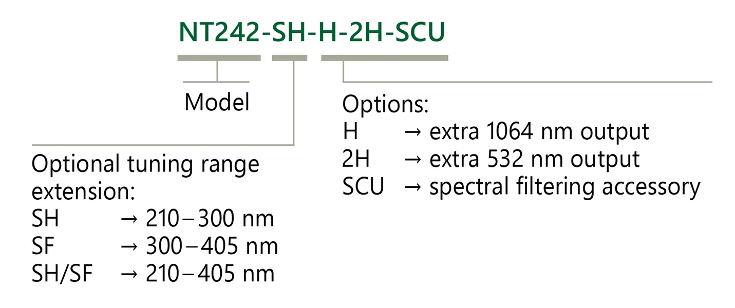
Ordering information of NT240 lasers.
Typical output pulse energy of NT242 series laser.

Typical beam profile of NT242 series lasers.
Near field at 500 nm.
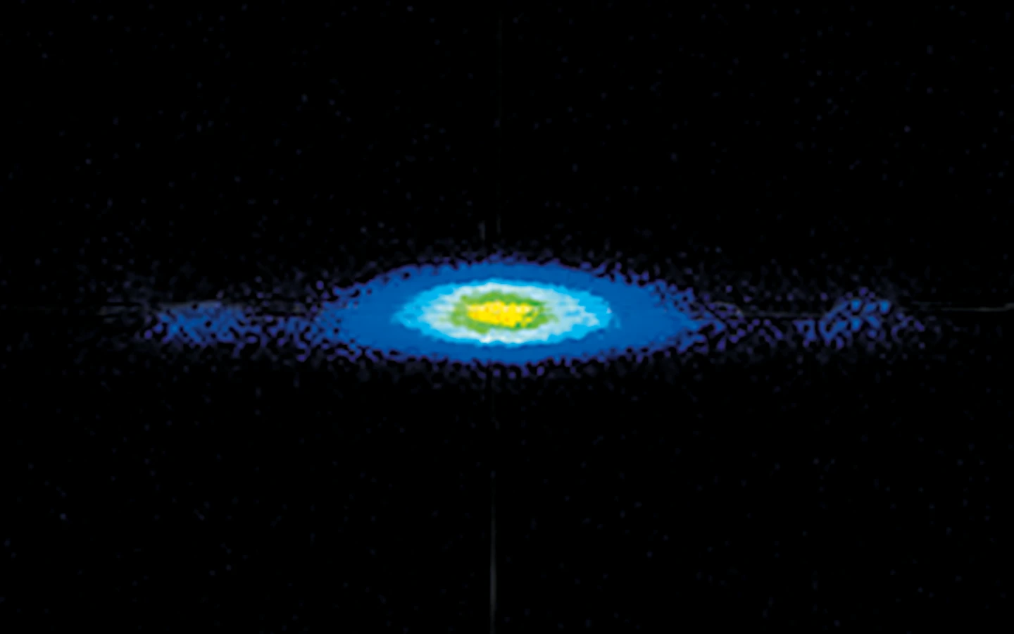
Typical beam profile of NT242 series lasers.
Far field at 500 nm.
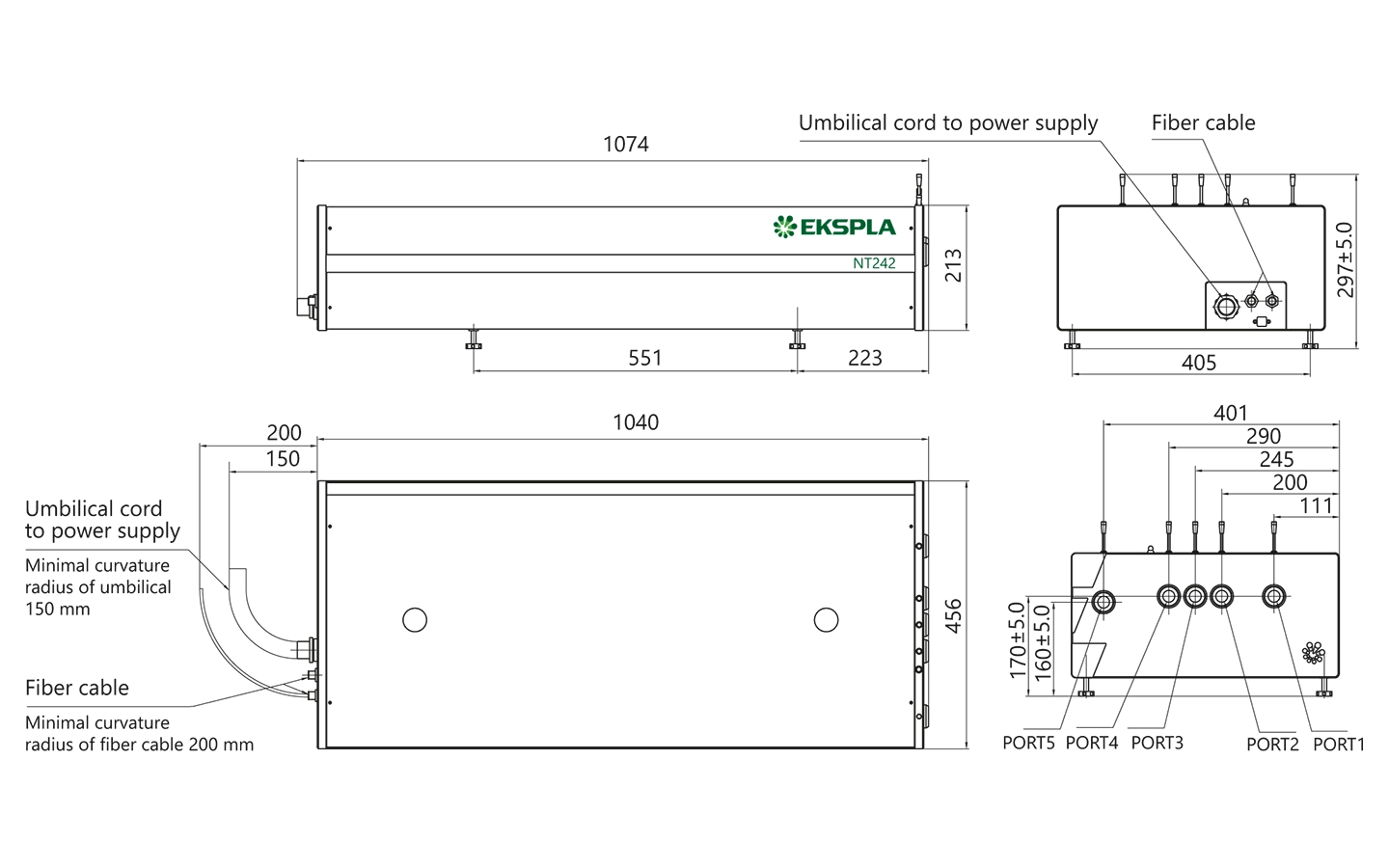
NT242 series laser head dimensions.
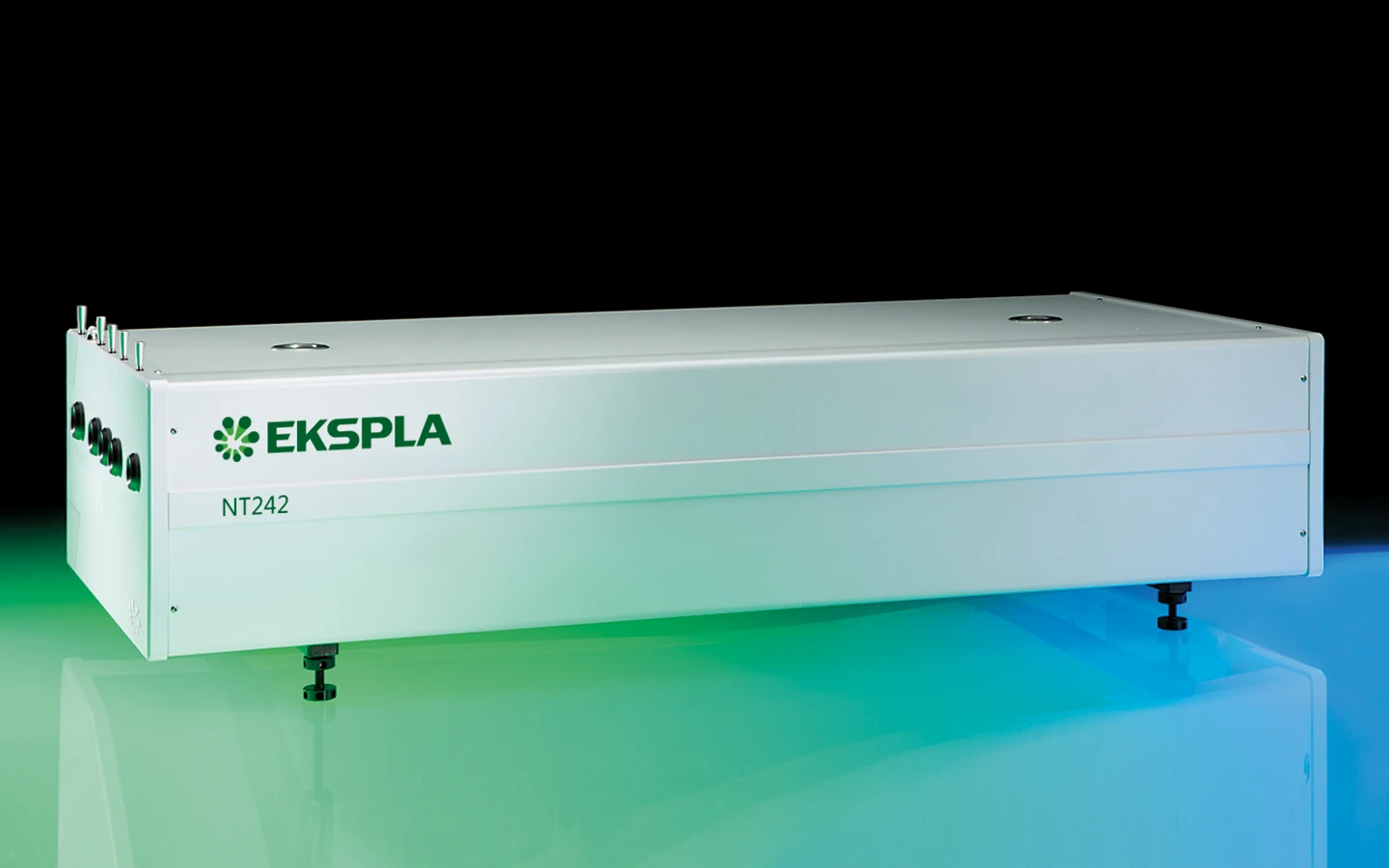
Broadly tunable kHz pulsed DPSS laser NT242.