SHG spectroscopy / microscopy
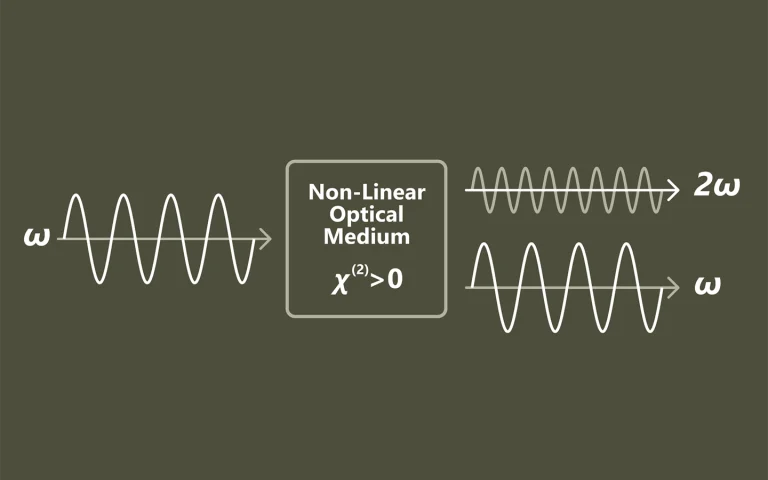
Second harmonic generation (SHG) is a second order nonlinear optical effect where two photons of frequency ω are converted to one photon of frequency 2ω. SHG is allowed only in media without inversion symmetry. This optical method it is non‐invasive, can be applied in situ, and can provide real time resolution.
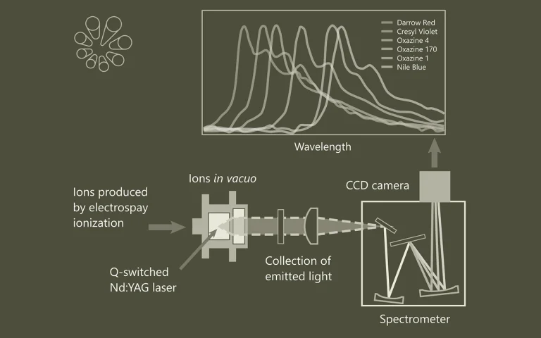
SHG measurements provide information about: surface coverage, molecular orientation, adsorbtion-desorbtion processes, and reactions at interfaces. SHG has the ability to detect low concentrations of analytes, such as proteins, peptides, and small molecules, due to its high sensitivity, and the second harmonic response can be enhanced through the use of target molecules that are resonant with the incident (ω) and/or second harmonic (2ω) frequencies.
SHG microscopy allows for selective probing of a non-centrosymmetric area of sample. This type of nonlinear optical microscope was first used to observe ferroelectric domains and has been applied to various specimens including the biological samples to date. Imaging of the endogenous SHG of biological tissue can be utilized for the selective observation of filament systems in tissues such as collagen, myosin, and microtubules, which exhibit a polar structure. It has been reported that, by imaging exogenous SHG of the membrane, sensitive detection of membrane damage could be realized using the SHG microscope.

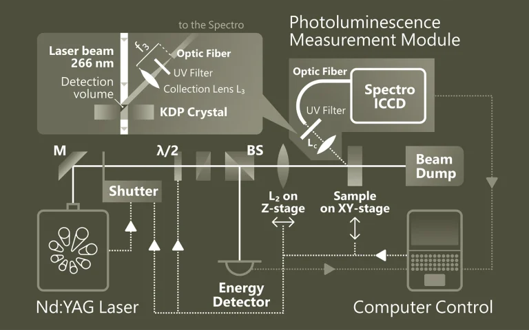
Principle of Second Harmonic Generation (SHG) Spectroscopy.