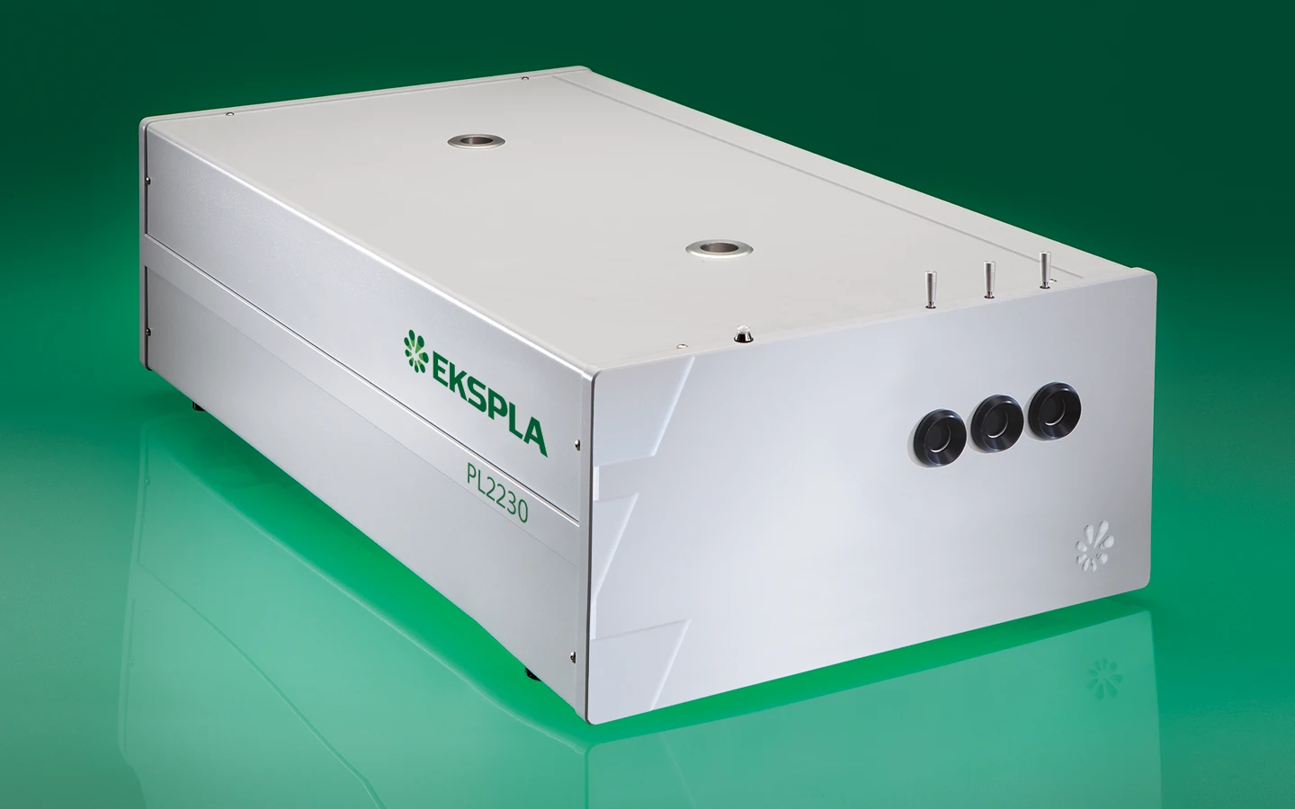
PL2230 series
Diode Pumped High Energy Picosecond Nd:YAG Lasers
PL2230 series diode-pumped, air-cooled, mode-locked picosecond Nd:YAG lasers provide high up to 40 mJ energy picosecond pulses at a 100 Hz pulse repetition rate. Precise pulse energy control, excellent short-term and long-term stabilit makes PL2230 series picosecond lasers an excellent choice for many demanding scientific applications.
Features
- Diode pumped power amplifier producing up to 40 mJ per pulse at 1064 nm
- Beam profile improvement using advanced beam shaping system
- Hermetically sealed DPSS master oscillator
- Diode pumped regenerative amplifier
- Air-cooled
- <30 ps pulse duration
- Excellent pulse duration stability
- Up to 100 Hz repetition rate
- Streak camera triggering pulse with <10 ps jitter
- Excellent beam pointing stability
- Thermo stabilized second, third or fourth harmonic generator options
- PC control
- Remote control via keypad
Applications
- Time resolved fluorescence (including streak camera measurements)
- SFG/SHG spectroscopy
- Nonlinear spectroscopy
- Laser-induced breakdown spectroscopy
- OPG pumping
- Remote laser sensing
- Satellite ranging
- Other spectroscopic and nonlinear optics applications
Description
Innovative design
The heart of the system is a diode pumped solid state (DPSS) master oscillator placed in a sealed monolithic block, producing high repetition rate pulse trains (90 MHz) with a low single pulse energy of several nJ. Diode pumped amplifiers are used for amplification of the pulse to 30 mJ or up to 40 mJ output. The high‑gain regenerative amplifier has an amplification factor in the proximity of 10⁶. After the regenerative amplifier, the pulse is directed to a multipass power amplifier that is optimized for efficient stored energy extraction from the Nd:YAG rod, while maintaining a near Gaussian beam profile and low wavefront distortion. The output pulse energy can be adjusted in approximately 1% steps, while pulse‑to-pulse energy stability remains at less than 0.5% rms at 1064 nm.
Angle-tuned KD*P and KDP crystals mounted in thermostabilised ovens are used for second, third, and fourth harmonic generation. Harmonic separators ensure the high spectral purity of each harmonic guided to different output ports.
Built-in energy monitors continuously monitor output pulse energy. Data from the energy monitor can be seen on the remote keypad or on a PC monitor. The laser provides triggering pulses for the synchronisation of your equipment. The lead of the triggering pulse can be up to 500 ns and is user adjustable in ~0.25 ns steps from a personal computer. Up to 1000 μs lead of triggering pulse is available as a pretrigger feature. Precise pulse energy control, excellent short-term and long-term stability, and a 50 Hz repetition rate makes PL2230 series lasers an excellent choice for many demanding scientific applications.
Simple and convenient laser control
For customer convenience the laser can be operated from master device or personal computer through USB (VCP, ASCII commands), RS232 (ASCII commands), LAN (REST API) or RS232 (ASCII commands), LAN (REST API) depending on the system configuration or from remote control pad with backlit display that is easy to read even while wearing laser safety glasses.
Communication interface | Description |
---|---|
USB | virtual serial port, ASCII commands |
RS232 | ASCII commands |
LAN | REST API |
WLAN | REST API |
Specifications
Model | PL2230-100 | PL2230A-100 | PL2231-50 | PL2231A-50 |
---|---|---|---|---|
Main specifications 1) | ||||
Pulse energy 2) | ||||
at 1064 nm | 3 mJ | 6 mJ | 30 mJ | 40 mJ |
at 532 nm 3) | 1.3 mJ | 3 mJ | 13 mJ | 18 mJ |
at 355 nm 4) | 0.9 mJ | 2 mJ | 9 mJ | 13 mJ |
at 266 nm 5) | 0.3 mJ | 0.6 mJ | 3 mJ | 5 mJ |
at 213 nm 6) | inquire | inquire | inquire | inquire |
Pulse energy stability (StdDev) 7) | ||||
at 1064 nm | < 0.2 % | < 0.6 % | < 0.5 % | < 0.5 % |
at 532 nm | < 0.4 % | < 0.4 % | < 0.8 % | < 0.8 % |
at 355 nm | < 0.5 % | < 0.5 % | < 1.1 % | < 1.1 % |
at 266 nm | < 0.5 % | < 0.5 % | < 1.2 % | < 1.2 % |
at 213 nm | < 1.5 % | < 1.5 % | < 1.5 % | < 1.5 % |
Pulse duration (FWHM) 8) | 29 ± 5 ps | 29 ± 5 ps | 29 ± 5 ps | 29 ± 5 ps |
Pulse duration stability 9) | ± 1 % | ± 1 % | ± 1 % | ± 1 % |
Power drift 10) | ± 2 % | ± 2 % | ± 2 % | ± 2 % |
Pulse repetition rate | ||||
at 1064, 532, 355 nm | 0 – 100 Hz | 100 Hz | 50 Hz | 50 Hz |
at 266, 213 nm | 100 Hz | 100 Hz | 10 Hz | 10 Hz |
Polarization 11) | vertical, > 99 % | vertical, > 99 % | vertical, > 99 % | vertical, > 99 % |
Pre-pulse contrast 12) | > 200 : 1 | > 200 : 1 | > 200 : 1 | > 200 : 1 |
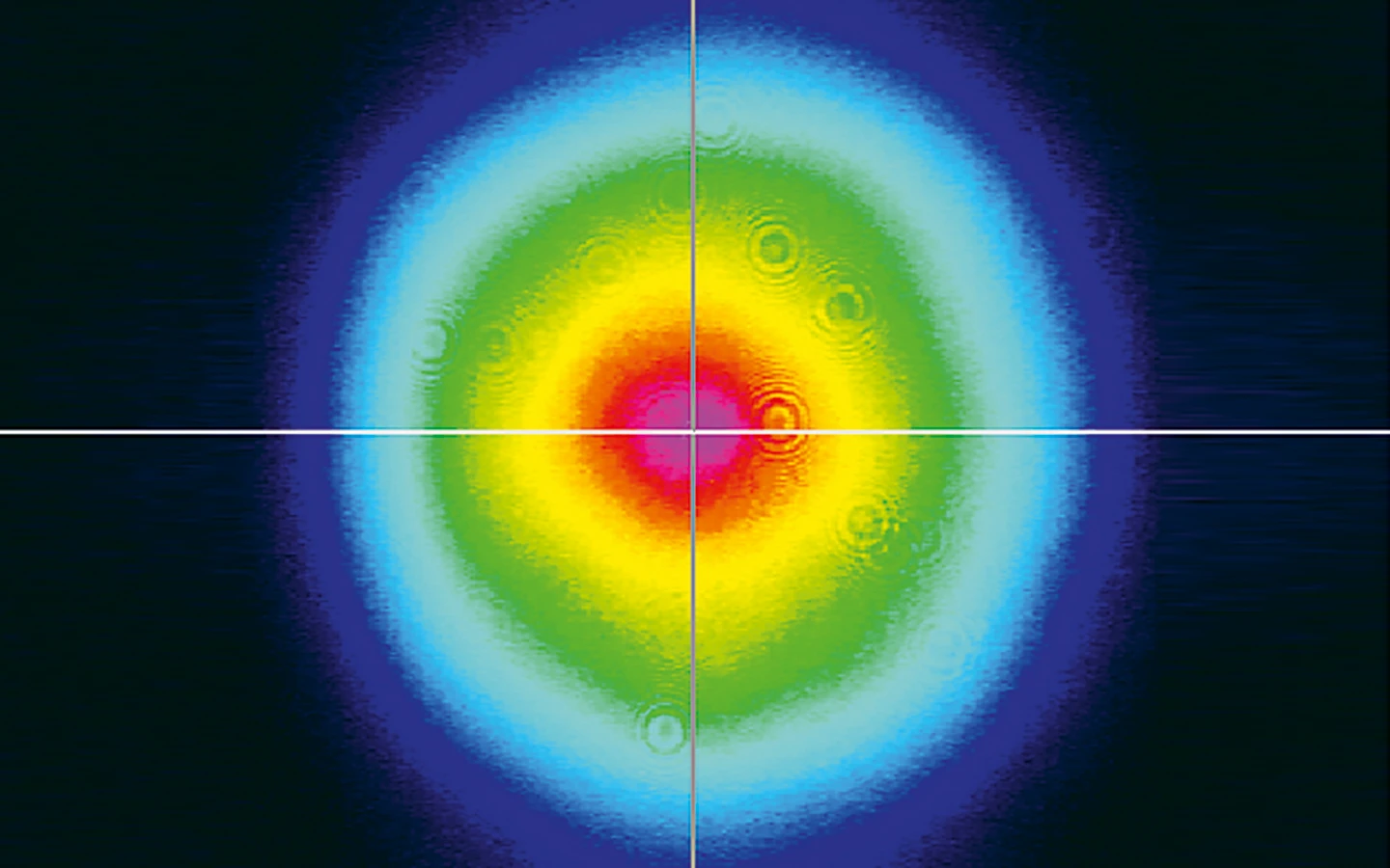
Beam profile 13) | close to Gaussian | close to Gaussian | close to Gaussian | close to Gaussian |
Beam divergence 14) | < 1.5 mrad | < 0.7 mrad | < 0.7 mrad | < 0.7 mrad |
Beam propagation ratio M2 | < 1.3 | < 1.3 | < 2.5 | < 2.5 |
Beam pointing stability (RMS) 15) | ≤ 10 μrad | ≤ 20 μrad | ≤ 20 μrad | ≤ 20 μrad |
Typical beam diameter 16) | ~ 2 mm | ~ 2.5 mm | ~ 6 mm | ~ 7 mm |
Optical pulse jitter | ||||
Internal triggering regime 17) | <50 ps | <50 ps | <50 ps | <50 ps |
External triggering regime 18) | ~3 ns | ~3 ns | ~3 ns | ~3 ns |
TRIG1 OUT pulse delay 19) | -500 … 50 ns | -500 … 50 ns | -500 … 50 ns | -500 … 50 ns |
Typical warm-up time | 5 min | 10 min | 15 min | 15 min |
Physical characteristics | ||||
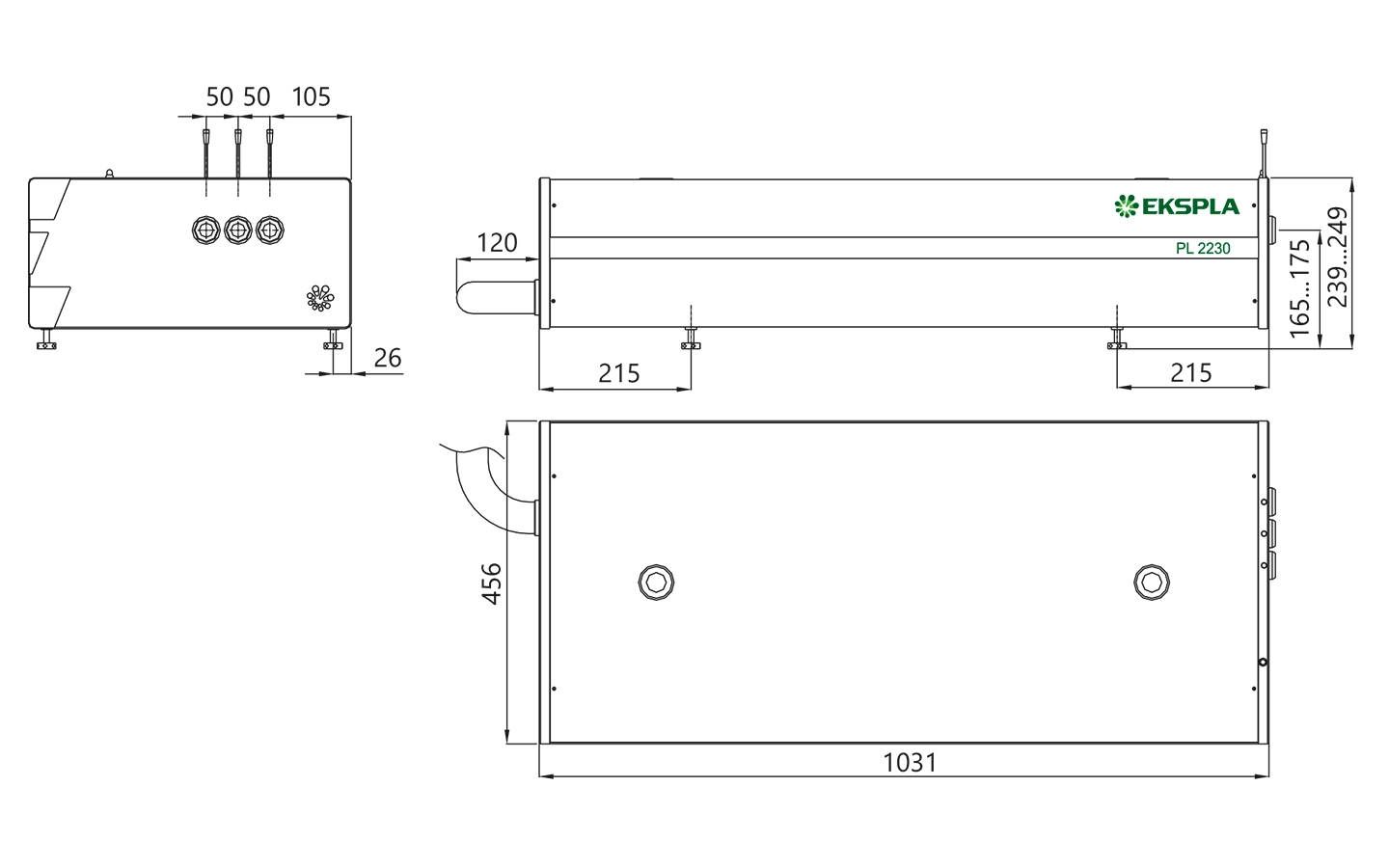
Laser head size (W × L × H) | 456×1031×249 ± 3 mm | 456×1031×249 ± 3 mm | 456×1031×249 ± 3 mm | 456×1031×249 ± 3 mm |
Electrical cabinet size (W × L × H) | 12 V DC power adapter, 85×170×41 ± 3 mm | 471×391×147 ± 3 mm | 471×391×147 ± 3 mm | 471×391×147 ± 3 mm |
Umbilical length | 2.5 m | 2.5 m | 2.5 m | 2.5 m |
Operating requirements | ||||
Cooling 20) | not required, air cooled | not required, air cooled | stand-alone chiller | stand-alone chiller |
Room temperature | 22 ± 2 °C | 22 ± 2 °C | 22 ± 2 °C | 22 ± 2 °C |
Relative humidity | 20 – 80 % (non-condensing) | 20 – 80 % (non-condensing) | 20 – 80 % (non-condensing) | 20 – 80 % (non-condensing) |
Power requirements | 110 – 240 V AC, 50/60 Hz | 110 – 240 V AC, 5 A, single phase 50/60 Hz | 110 – 240 V AC, 5 A, single phase 50/60 Hz | 110 – 240 V AC, 5 A, single phase 50/60 Hz |
Power consumption | < 0.15 kVA | < 1.0 kVA | < 1.0 kVA | < 1.0 kVA |
Model | PL2230-100 | PL2230A-100 | PL2231-50 | PL2231A-50 |
---|
- Due to continuous improvement, all specifications are subject to change without notice. Parameters marked typical are not specifications. They are indications of typical performance and will vary with each unit we manufacture. Unless stated otherwise, all specifications are measured at 1064 nm and for basic system without options.
- Outputs are not simultaneous.
- For PL2230 series laser with –SH, -SH/TH, -SH/FH or -SH/TH/FH option or –SH/TH/FH/FiH module.
- For PL2230 series laser with –TH, -SH/TH or -SH/TH/FH option or –SH/TH/FH/FiH module.
- For PL2230 series laser with -SH/FH or -SH/TH/FH option or –SH/TH/FH/FiH module.
- For PL2230 series laser with –SH/TH/FH/FiH module.
- Averaged from pulses, emitted during 30 sec time interval.
- FWHM. Inquire for optional pulse durations in 20 – 90 ps range. Pulse energy specifications may differ from indicated here.
- Measured over 1 hour period when ambient temperature variation is less than ±1 °C.
- Measured over 8 hours period after 20 min warm-up when ambient temperature variation is less than ± 2 °C.
- At 1064 nm.
- Peak-to-peak with respect to residual pulses.
- In near and far fields. Near field Gaussian fit is >80%.
- Average of X- and Y-plane full angle divergence values measured at the 1/e2 level at 1064 nm.
- Beam pointing stability is evaluated from fluctuations of beam centroid position in the far field.
- Beam diameter is measured at 1064 nm at the 1/e2 level.
- StdDev. With respect to TRIG1 OUT pulse. <10 ps jitter is provided optionally with PRETRIG feature.
- StdDev. With respect to SYNC IN pulse.
- TRIG1 OUT lead or delay can be adjusted with 0.25 ns steps in specified range.
- Air cooled. Adequate room air conditioning should be provided.
Note: Laser must be connected to the mains electricity all the time. If there will be no mains electricity for longer that 1 hour then laser (system) needs warm up for a few hours before switching on.
Options
-P20
Provides 20 ps ±10% output pulse duration. Pulse energies are ~ 30 % lower in comparison to the 29 ps pulse duration version. See table below for pulse energy specifications:
Model | PL2231-50 | PL2231A-50 |
---|---|---|
1064 nm | 23 mJ | 28 mJ |
532 nm | 9 mJ | 13 mJ |
355 nm | 6 mJ | 9 mJ |
266 nm | 2 mJ | 4 mJ |
PL2231A-50 HE
Pulse repetition rate 50 Hz. The pulse energy is ~30% higher compared to the laser without depolarization compensation. 29±5 ps output pulse duration. See table below for pulse energy specifications:
Model 1) 2) | PL2231A-50 HE |
---|---|
1064 nm | 50 mJ |
- Due to continuous improvement, all specifications are subject to change without notice. Parameters marked typical are not specifications. They are indications of typical performance and will vary with each unit we manufacture. Unless stated otherwise, all specifications are measured at 1064 nm and for basic system without options. Specifications for model PL2231C are preliminary and should be confirmed against quotation and purchase order.
- Outputs are not simultaneous.
-P80
Provides 80 ps ± 10% output pulse duration. Pulse energy specifications are same as those of 29 ps lasers.
-P10
10±2 ps pulse duration. Pulse energies are ~ 50% lower in comparison to the 29 ps pulse duration version. Valid only for PL2230A-100.
-PLL
Allows locking the master oscillator pulse train repetition rate to an external RF generator, enabling precise external triggering with low jitter. Inquire for more information.
PL2231A-10
Pulse repetition rate 10 Hz. The pulse energy is ~2 times higher compared to the 50 Hz laser version. 29±5 ps output pulse duration. See table below for pulse energy specifications:
Model 1) 2) | PL2231A-10 |
---|---|
1064 nm | 80 mJ |
532 nm 3) | 50 mJ |
355 nm | inquire |
216 nm | inquire |
213 nm | inquire |
- Due to continuous improvement, all specifications are subject to change without notice. Parameters marked typical are not specifications. They are indications of typical performance and will vary with each unit we manufacture. Unless stated otherwise, all specifications are measured at 1064 nm and for basic system without options. Specifications for model PL2231C are preliminary and should be confirmed against quotation and purchase order.
- Outputs are not simultaneous.
- For PL2231A-10 series laser with –SH module.
Performance and drawings
Publications
Ultrafast transient absorption spectra and kinetics of human blue cone visual pigment at room temperature
The ultrafast photochemical reaction mechanism, transient spectra, and transition kinetics of the human blue cone visual pigment have been recorded at room temperature. Ultrafast time-resolved absorption spectroscopy revealed the progressive formation and decay of several metastable photo-intermediates, corresponding to the Batho to Meta-II photo-intermediates previously observed with bovine rhodopsin and human green cone opsin, on the picosecond to millisecond timescales following pulsed excitation. The experimental data reveal several interesting similarities and differences between the photobleaching sequences of bovine rhodopsin, human green cone opsin, and human blue cone opsin. While Meta-II formation kinetics are comparable between bovine rhodopsin and blue cone opsin, the transition kinetics of earlier photo-intermediates and qualitative characteristics of the Meta-I to Meta-II transition are more similar for blue cone opsin and green cone opsin. Additionally, the blue cone photo-intermediate spectra exhibit a high degree of overlap with uniquely small spectral shifts. The observed variation in Meta-II formation kinetics between rod and cone visual pigments is explained based on key structural differences.
Near infrared-triggered liposome cages for rapid, localized small molecule delivery
Photolabile chelating cages or protecting groups need complex chemical syntheses and require UV, visible, or two-photon NIR light to trigger release. Different cages have different solubilities, reaction rates, and energies required for triggering. Here we show that liposomes containing calcium, adenosine triphosphate, or carboxyfluorescein are tethered to plasmon-resonant hollow gold nanoshells (HGN) tuned to absorb light from 650–950 nm. Picosecond pulses of near infrared (NIR) light provided by a two-photon microscope, or by a stand-alone laser during flow through microfluidic channels, trigger contents release with spatial and temporal control. NIR light adsorption heats the HGN, inducing vapor nanobubbles that rupture the liposome, releasing cargo within milliseconds. Any water-soluble molecule can be released at essentially the same rate from the liposome-HGN. By using liposomes of different composition, or HGN of different sizes or shapes with different nanobubble threshold fluences, or irradiating on or off resonance, two different cargoes can be released simultaneously, one before the other, or in a desired ratio. Calcium release from liposome-HGN can be spatially patterned to crosslink alginate gels and trap living cells. Liposome-HGN provide stable, biocompatible isolation of the bioactive compound from its surroundings with minimal interactions with the local environment.
Structure Determination of Hen Egg-White Lysozyme Aggregates Adsorbed to Lipid/Water and Air/Water Interfaces
We use vibrational sum-frequency generation (VSFG) spectroscopy to study the structure of hen egg-white lysozyme (HEWL) aggregates adsorbed to DOPG/D2O and air/D2O interfaces. We find that aggregates with a parallel and antiparallel β-sheet structure together with smaller unordered aggregates and a denaturated protein are adsorbed to both interfaces. We demonstrate that to retrieve this information, fitting of the VSFG spectra is essential. The number of bands contributing to the VSFG spectrum might be misinterpreted, due to interference between peaks with opposite orientation and a nonresonant background. Our study identified hydrophobicity as the main driving force for adsorption to the air/D2O interface. Adsorption to the DOPG/D2O interface is also influenced by hydrophobic interaction; however, electrostatic interaction between the charged protein’s groups and the lipid’s headgroups has the most significant effect on the adsorption. We find that the intensity of the VSFG spectrum at the DOPG/D2O interface is strongly enhanced by varying the pH of the solution. We show that this change is not due to a change of lysozyme’s and its aggregates’ charge but due to dipole reorientation at the DOPG/D2O interface. This finding suggests that extra care must be taken when interpreting the VSFG spectrum of proteins adsorbed at the lipid/water interface.
A primary radiation standard based on quantum nonlinear optics
The black body remains the most prominent source of light for absolute radiometry. Its main alternative, synchrotron radiation, requires costly and large facilities. Quantum optics offers a new radiometric source: parametric down-conversion (PDC), a nonlinear optical process, in which pairwise photon correlations enable absolute calibration of photodetectors. Since the emission rate crucially depends on the brightness of the electromagnetic field, quantum-mechanical fluctuations of the vacuum can be seen as a seed of spontaneous PDC, and their amplitude is a natural radiometric standard. Thus, they allow for the calibration of the spectral radiance of light sources by measuring the ratio between seeded and unseeded PDC. Here, we directly use the frequency spectrum of the electromagnetic vacuum to trigger spontaneous PDC and employ the generated light to infer the spectral response of a spectrometer over a broad spectral range. Then, we deduce the absolute quantum efficiency from the spectral shape of PDC in the high-gain regime, without relying on a seed or reference detector. Our results compare well with the ones obtained with a reference lamp, demonstrating a promising primary radiation standard.
High-performance all-organic DFB and DBR waveguide laser with various grating height fabricated by a two-photon absorption DLW method
Organic solid-state lasers (OSSLs) with distributed feedback (DFB) structures or distributed Bragg reflectors (DBRs) are promising for potential application in bio-sensing and hazardous materials detection. Here, the laser performances of the all-organic DFB waveguide lasers with various grating heights ranging from 0.4 to 4.7 μm were investigated. The grating structures used as the lasing cavity were fabricated using a two-photon absorption (TPA) direct laser writing (DLW) method with an SU-8 negative photoresist. The laser active layer consisted of a rhodamine 6G (R6G) laser dye and a cellulose acetate (CA) matrix. The R6G/CA solution was spin-coated onto the quartz substrate with the cavity (grating) structures to fabricate the DFB waveguide laser devices. The diffraction order of lasing ranged from m = 4 to 7. As the grating height was increased to 1.9 μm, the slope efficiency increased for all diffraction orders and the threshold decreases for each diffraction order. The dependence of the cavity (grating) length on the laser performances was investigated. The slope efficiency increased as the cavity length increased to 300 μm. The effect of the cavity (grating) position on the slope efficiency and the threshold position of the cavity (grating) was also studied. A maximum slope efficiency of 10.2% was achieved for the DFB waveguide laser device with a cavity (grating) length of 300 μm, a cavity position at 6 mm from the emission edge of the waveguide, and an aspect ratio ≈3 between the grating height of 1.74 μm and the grating width of 0.6 μm for the diffraction order m = 6 for lasing.
Vibrational Relaxation Lifetime of a Physisorbed Molecule at a Metal Surface
Previous measurements of vibrational relaxation lifetimes for molecules adsorbed at metal surfaces yielded values of 1–3 ps; however, only chemisorbed molecules have been studied. We report the first measurements of the vibrational relaxation lifetime of a molecule physisorbed to a metal surface. For CO(υ=1) adsorbed on Au(111) at 35 K the vibrational lifetime of the excited stretching mode is 49±3 ps. The long lifetime seen here is likely to be a general feature of physisorption, which involves weaker electronic coupling between the adsorbate and the solid due to bonding at larger distances.
Heavy Anionic Complex Creates a Unique Water Structure at a Soft Charged Interface
Ion hydration and interfacial water play crucial roles in numerous phenomena ranging from biological to industrial systems. Although biologically relevant (and mostly smaller) ions have been studied extensively in this context, very little experimental data exist about molecular-scale behavior of heavy ions and their complexes at interfaces, especially under technologically significant conditions. It has recently been shown that PtCl62– complexes adsorb at positively charged interfaces in a two-step process that cannot fit into well-known empirical trends, such as Hofmeister series. Here, a combined vibrational sum frequency generation and molecular dynamics study reveals that a unique interfacial water structure is connected to this peculiar adsorption behavior. A novel subensemble analysis of molecular dynamics simulation results shows that after adsorption PtCl62– complexes partially retain their first and second hydration spheres and that it is possible to identify three different types of water molecules around them on the basis of their orientational structures and hydrogen-bonding strengths. These results have important implications for relating interfacial water structure and hydration enthalpy to the general understanding of specific ion effects. This in turn influences interpretation of heavy metal ion distribution across, and reactivity within, liquid interfaces.
How nature covers its bases
The response of DNA and RNA bases to ultraviolet (UV) radiation has been receiving increasing attention for a number of important reasons: (i) the selection of the building blocks of life on an early earth may have been mediated by UV photochemistry, (ii) radiative damage of DNA depends critically on its photochemical properties, and (iii) the processes involved are quite general and play a role in more biomolecules as well as in other compounds. A growing number of groups worldwide have been studying the photochemistry of nucleobases and their derivatives. Here we focus on gas phase studies, which (i) reveal intrinsic properties distinct from effects from the molecular environment, (ii) allow for the most detailed comparison with the highest levels of computational theory, and (iii) provide isomeric selectivity. From the work so far a picture is emerging of rapid decay pathways following UV excitation. The main understanding, which is now well established, is that canonical nucleobases, when absorbing UV radiation, tend to eliminate the resulting electronic excitation by internal conversion (IC) to the electronic ground state in picoseconds or less. The availability of this rapid “safe” de-excitation pathway turns out to depend exquisitely on molecular structure. The canonical DNA and RNA bases are generally short-lived in the excited state, and thus UV protected. Many closely related compounds are longer lived, and thus more prone to other, potentially harmful, photochemical processes. It is this structure dependence that suggests a mechanism for the chemical selection of the building blocks of life on an early earth. However, the picture is far from complete and many new questions now arise.
Ultra-sensitive mid-infrared emission spectrometer with sub-ns temporal resolution
We evaluate the performance of a mid-infrared emission spectrometer operating at wavelengths between 1.5 and 6 μm based on an amorphous tungsten silicide (a-WSi) superconducting nanowire single-photon detector (SNSPD). We performed laser induced fluorescence spectroscopy of surface adsorbates with sub-monolayer sensitivity and sub-nanosecond temporal resolution. We discuss possible future improvements of the SNSPD-based infrared emission spectrometer and its potential applications in molecular science.
Vibrational fingerprint of localized excitons in a two-dimensional metal-organic crystal
Long-lived excitons formed upon visible light absorption play an essential role in photovoltaics, photocatalysis, and even in high-density information storage. Here, we describe a self-assembled two-dimensional metal-organic crystal, composed of graphene-supported macrocycles, each hosting a single FeN4 center, where a single carbon monoxide molecule can adsorb. In this heme-like biomimetic model system, excitons are generated by visible laser light upon a spin transition associated with the layer 2D crystallinity, and are simultaneously detected via the carbon monoxide ligand stretching mode at room temperature and near-ambient pressure. The proposed mechanism is supported by the results of infrared and time-resolved pump-probe spectroscopies, and by ab initio theoretical methods, opening a path towards the handling of exciton dynamics on 2D biomimetic crystals.