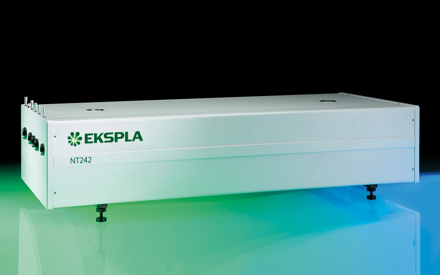
NT240 series
Broadly Tunable kHz Pulsed DPSS Lasers
NT240 delivers hands‑free, no-gap tuning from 210 to 2600 nm from the one box. With its 1000 Hz repetition rate, the NT240 series laser establishes itself as a versatile tool for many laboratory applications, including laser induced fluorescence, flash photolysis, photobiology, metrology, remote sensing.
Features
- Customers recognized reliability
- Two years warranty
- Integrates DPSS pump laser and OPO into a single housing
- Hands-free no-gap wavelength tuning from 210 to 2600 nm*
- 1000 Hz pulse repetition rate
- More than 60 µJ output pulse energy in UV
- Less than 5 cm‑1 linewidth
- 3 – 6 ns pulse duration
- Remote control via key pad or PC
- Optional separate output for the OPO pump beam 355 nm, 532 nm or 1064 nm
* Automatic wavelength scan is programmable
Applications
- Laser-induced fluorescence spectroscopy
- Pump-probe spectroscopy
- Non-linear spectroscopy
- Time-resolved spectroscopy
- Photobiology
- Remote sensing
- Determination of the telescope throughput
Description
NT240 series lasers produce pulses at an unprecedented 1 kHz pulse repetition rate, tunable over a broad spectral range.
Integrated into a single compact housing, the diode pumped Q-switched Nd:YAG laser and OPO offers hands‑free, no-gap tuning from 210 to 2600 nm. With its 1000 Hz repetition rate, the NT240 series laser establishes itself as a versatile tool for many laboratory applications, including laser induced fluorescence, flash photolysis, photobiology, metrology, remote sensing, etc.
NT240 series systems can be controlled from a remote control pad or/and a computer using supplied LabVIEW™ drivers. The control pad allows easy control of all parameters and features on a backlit display that is easy to read even with laser safety eyewear.
Thanks to a DPSS pump source, the laser requires little maintenance. It is equipped with air-cooled built-in chiller, which further reduces running costs. A built‑in OPO pump energy monitor allows monitoring of pump laser performance without the use of external power meters. The optional feature provides a separate output port for the 1064, 532 or 355 nm beam.
Benefits
- Hands-free wavelength tuning – no need for physical intervention
- High repetition rate 1000 Hz enables fast data collection
- End pumping with diode technology ensures high reliability and low maintenance costs
- Narrow linewidth (down to 3 cm‑1) and superior tuning resolution
- (1 – 2 cm‑1) allow recording of high quality spectra
- High integration level saves valuable space in the laboratory
- In-house design and manufacturing of complete systems, including pump lasers, guarantees on-time warranty and post warranty services and spares supply
- Variety of control interfaces: USB, RS232, LAN and WLAN ensures easy control and integration with other equipment
- Attenuator and fiber coupling options facilitate incorporation of NT240 systems into various experimental environments
Specifications
| Model | NT242 | NT242-SH | NT242-SF | NT242-SH/SF |
|---|---|---|---|---|
| OPO specifications 1) | ||||
| Wavelength range | ||||
| Signal | 405 – 710 nm | 405 – 710 nm | 405 – 710 nm | 405 – 710 nm |
| Idler | 710 – 2600 nm | 710 – 2600 nm | 710 – 2600 nm | 710 – 2600 nm |
| SH and SF | — | 210 – 300 nm | 300 – 405 nm | 210 – 405 nm |
| Pulse energy 2) | ||||
| OPO | 450 μJ | 450 μJ | 450 μJ | 450 μJ |
| SH and SF | — | 40 μJ at peak | 60 μJ at peak | 60 μJ at peak |
| Pulse repetition rate | 1000 Hz | 1000 Hz | 1000 Hz | 1000 Hz |
| Pulse duration 3) | 3 – 6 ns | 3 – 6 ns | 3 – 6 ns | 3 – 6 ns |
| Linewidth 4) | < 5 cm‑1 | < 5 cm‑1 | < 5 cm‑1 | < 5 cm‑1 |
| Minimal tuning step 5) | ||||
| Signal | 1 cm‑1 | 1 cm‑1 | 1 cm‑1 | 1 cm‑1 |
| Idler | 1 cm‑1 | 1 cm‑1 | 1 cm‑1 | 1 cm‑1 |
| SH and SF | — | 2 cm‑1 | 2 cm‑1 | 2 cm‑1 |
| Polarization | ||||
| Signal | horizontal | horizontal | horizontal | horizontal |
| Idler | vertical | vertical | vertical | vertical |
| SH and SF | — | vertical | vertical | vertical |
| Typical beam diameter 6) | 3 × 6 mm | 3 × 6 mm | 3 × 6 mm | 3 × 6 mm |
| Pump laser | ||||
| Pump wavelength 7) | 355 nm | 355 nm | 355 / 1064 nm | 355 / 1064 nm |
| Typical pump pulse energy 8) | 3 mJ | 3 mJ | 3 / 1 mJ | 3 / 1 mJ |
| Pulse duration 3) | 4 – 6 ns at 1064 nm | 4 – 6 ns at 1064 nm | 4 – 6 ns at 1064 nm | 4 – 6 ns at 1064 nm |
| Physical characteristics | ||||
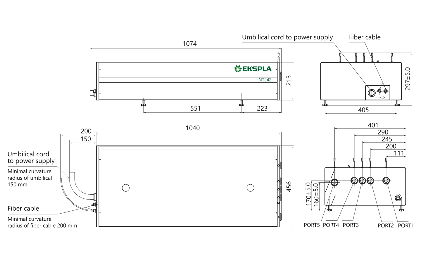
| Unit size (W × L × H) 9) | 456 × 1040 × 297 mm | 456 × 1040 × 297 mm | 456 × 1040 × 297 mm | 456 × 1040 × 297 mm |
| Power supply size (W × L × H) | 520 × 400 × 286 mm | 520 × 400 × 286 mm | 520 × 400 × 286 mm | 520 × 400 × 286 mm |
| Umbilical length | 2.5 m | 2.5 m | 2.5 m | 2.5 m |
| Operating requirements | ||||
| Cooling | built-in chiller | built-in chiller | built-in chiller | built-in chiller |
| Room temperature | 18 – 27 °C | 18 – 27 °C | 18 – 27 °C | 18 – 27 °C |
| Relative humidity | 20 – 80 % (non-condensing) | 20 – 80 % (non-condensing) | 20 – 80 % (non-condensing) | 20 – 80 % (non-condensing) |
| Power requirements | 100 – 240 V AC, single phase, 50/60 Hz | 100 – 240 V AC, single phase, 50/60 Hz | 100 – 240 V AC, single phase, 50/60 Hz | 100 – 240 V AC, single phase, 50/60 Hz |
| Power consumption | < 1.5 kW | < 1.5 kW | < 1.5 kW | < 1.5 kW |
| Cleanliness of the room | not worse than ISO Class 9 | not worse than ISO Class 9 | not worse than ISO Class 9 | not worse than ISO Class 9 |
| Model | NT242 | NT242-SH | NT242-SF | NT242-SH/SF |
|---|
- Due to continuous improvement, all specifications are subject to change. Parameters marked typical are illustrative; they are indications of typical performance and will vary with each unit we manufacture. Unless stated otherwise, all specifications are measured at 450 nm and for basic system without options.
- See tuning curves for typical outputs at other wavelengths.
- Measured at FWHM level with photodiode featuring 1 ns rise time and 300 MHz bandwidth oscilloscope.
- Linewidth is <8 cm‑1 for 210 – 405 nm range.
- For manual input from PC. When wavelength is controlled from keypad, tuning resolution is 0.1 nm for signal, 1 nm for idler and 0.05 nm for SH and SF.
- Beam diameter is measured at 450 nm at the 1/e2 level and can vary depending on the pump pulse energy.
- Separate output port for the 3rd and other harmonic is optional.
- The pump laser pulse energy will be optimized for best OPO performance. The actual pump laser output can vary with each unit we manufacture.
- Length from 1040 to 1233 mm depending on configuration.
Note: Laser must be connected to the mains electricity all the time. If there will be no mains electricity for longer that 1 hour then laser (system) needs warm up for a few hours before switching on.
Accessories and optional items
| Option | Features |
|---|---|
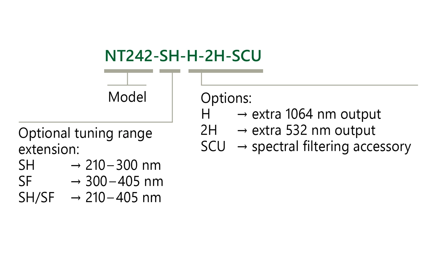
| -SH | Tuning range extension in UV range (210 – 300 nm) by second harmonic generation |
| -SF | Tuning range extension in 300 – 405 nm range by sum-frequency generation |
| -SH/SF | Tuning range extension in 210 – 405 nm range by combining second harmonics and sum-frequency generator outputs for maximum possible pulse energy |
| -SCU | Spectral filtering accessory for improved spectral purity of pulses |
| -H, -2H, -3H | 1064, 532 and 355 nm output via separate port |
| -FC | Fiber coupler |
| -ATTN | Attenuator option |
| Option | Features |
|---|
Performance

Typical output pulse energy of NT242 series tunable laser.
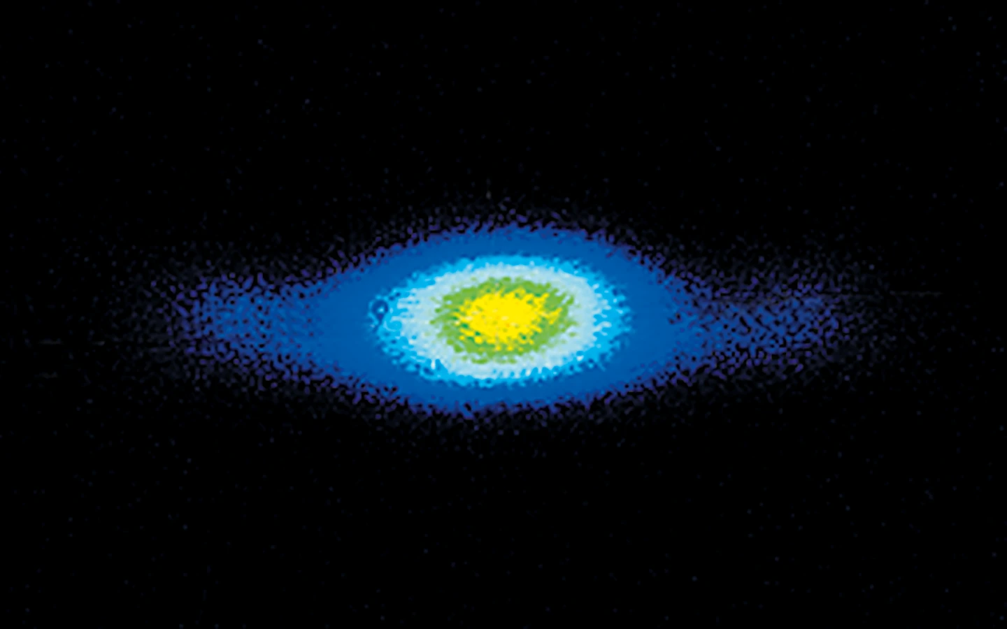
Typical beam profiles of NT242 series lasers at 500 nm. Near field.
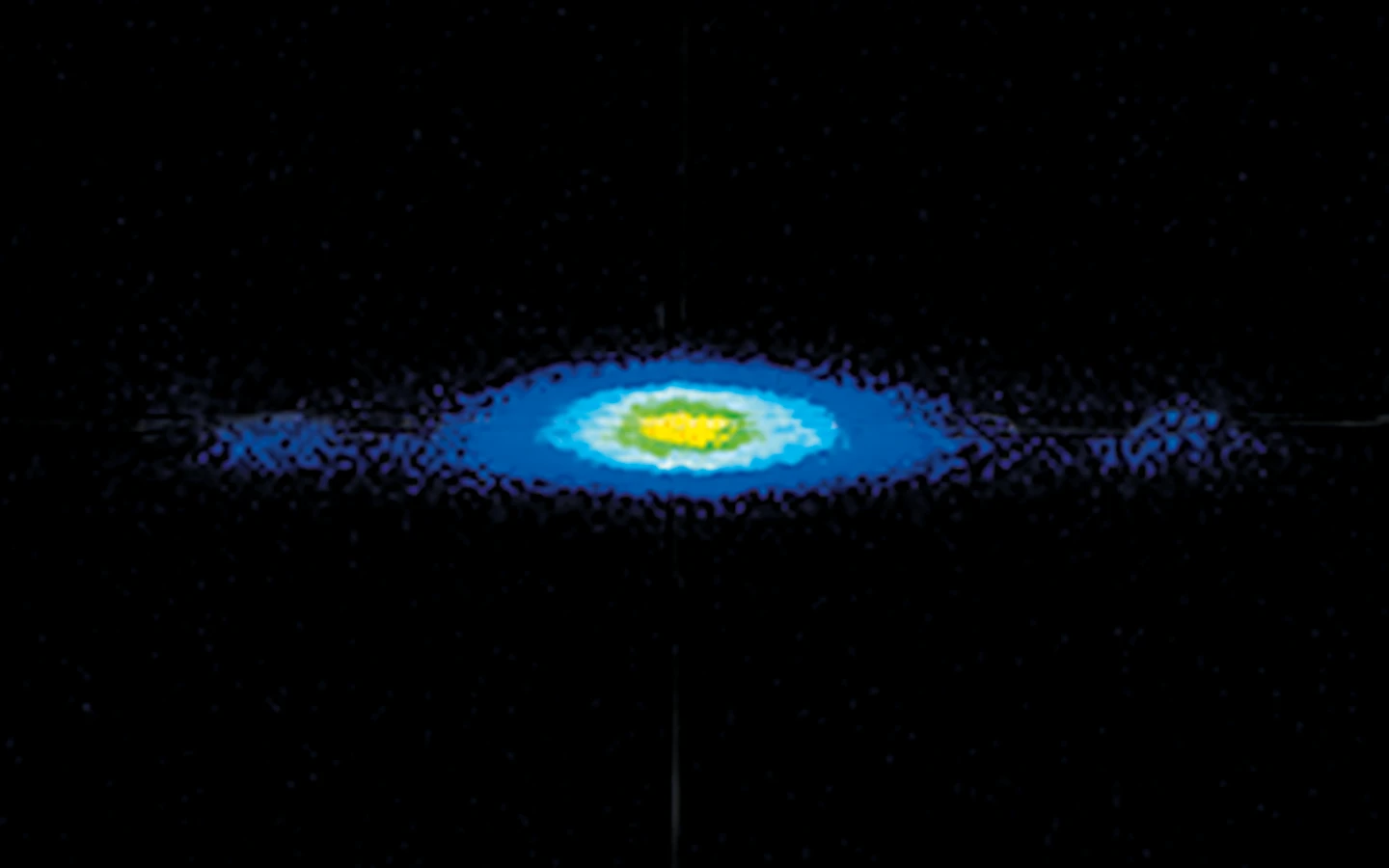
Typical beam profiles of NT242 series lasers at 500 nm. Far field.
Publications
Considerable matrix shift in the electronic transitions of helium-solvated cesium dimer cation Cs2He+n
We investigate the photodissociation of helium-solvated cesium dimer cations using action spectroscopy and quantum chemical calculations. The spectrum of Cs2He+ shows three distinct absorption bands into both bound and dissociative states. Upon solvation with further helium atoms, considerable shifts of the absorption bands are observed, exceeding 0.1 eV (850 cm−1) already for Cs2He10+, along with significant broadening. The shifts are highly sensitive to the character of the excited state. Our calculations show that helium atoms adsorb on the ends of Cs2+. The shifts are particularly pronounced if the excited state orbitals extend to the area occupied by the helium atoms. In this case, Pauli repulsion leads to a deformation of the excited state orbitals, resulting in the observed blue shift of the transition. Since the position of the weakly bound helium atoms is ill defined, Pauli repulsion also explains the broadening.
Contrast agent enhanced multimodal photoacoustic microscopy and optical coherence tomography for imaging of rabbit choroidal and retinal vessels in vivo
Multimodal imaging with photoacoustic microscopy (PAM) and optical coherence tomography (OCT) can be an effective method to evaluate the choroidal and retinal microvasculature. To improve the efficiency for visualizing capillaries, colloidal gold nanoparticles (AuNPs) have been applied as a multimodal contrast agent for both OCT and PAM imaging by taking advantage of the strong optical scattering and the strong optical absorption of AuNPs due to their surface plasmon resonance. Ultra-pure AuNPs were fabricated by femtosecond laser ablation, capped with polyethylene glycol (PEG), and administered to 13 New Zealand white rabbits and 3 Dutch Belted pigmented rabbits. The synthesized PEG-AuNPs (20.0 ± 1.5 nm) were demonstrated to be excellent contrast agents for PAM and OCT, and do not demonstrate cytotoxicity to bovine retinal endothelial cells in cell studies. The image signal from the retinal and choroidal vessels in living rabbits was enhanced by up to 82% for PAM and up to 45% for OCT, respectively, by the administered PEG-AuNPs, which enables detection of individual blood vessels by both imaging modalities. The biodistribution study demonstrated the AuNP accumulated primarily in the liver and spleen. Histology and TUNEL staining did not indicate cell injury or death in the lung, liver, kidney, spleen, heart, or eyes up to seven days after AuNP administration. PEG-AuNPs offer an efficient and safe contrast agent for multimodal ocular imaging to achieve improved characterization of microvasculature.
Electronic spectroscopy and nanocalorimetry of hydrated magnesium ions [Mg(H2O)n]+, n = 20 – 70: spontaneous formation of a hydrated electron?
Hydrated singly charged magnesium ions [Mg(H2O)n]+ are thought to consist of an Mg2+ ion and a hydrated electron for n > 15. This idea is based on mass spectra, which exhibit a transition from [MgOH(H2O)n−1]+ to [Mg(H2O)n]+ around n = 15 – 22, black-body infrared radiative dissociation, and quantum chemical calculations. Here, we present photodissociation spectra of size-selected [Mg(H2O)n]+ in the range of n = 20 – 70 measured for photon energies of 1.0 – 5.0 eV. The spectra exhibit a broad absorption from 1.4 to 3.2 eV, with two local maxima around 1.7 – 1.8 eV and 2.1 – 2.5 eV, depending on cluster size. The spectra shift slowly from n = 20 to n = 50, but no significant change is observed for n = 50 – 70. Quantum chemical modeling of the spectra yields several candidates for the observed absorptions, including five- and six-fold coordinated Mg2+ with a hydrated electron in its immediate vicinity, as well as a solvent-separated Mg2+/e− pair. The photochemical behavior resembles that of the hydrated electron, with barrierless interconversion into the ground state following the excitation.
High-resolution multimodal photoacoustic microscopy and optical coherence tomography image-guided laser induced branch retinal vein occlusion in living rabbits
Joint high-resolution multimodal photoacoustic microscopy (PAM) and optical coherence tomography (OCT) was developed to improve the efficiency for visualizing newly developed retinal neovascularization (RNV) and to monitor the dynamic changes of retinal vein occlusion (RVO) in living rabbits. The RNV and RVO models were created in New Zealand rabbits by Rose Bengal laser-induced RVO. Dual modalities imaging equipment, including color fundus photography, fluorescein angiography (FA), OCT, and PAM, was used to image and assess the changes of retinal vasculature. In vivo experimental results exhibited that not only the treatment boundaries and the position of the occluded vasculature but also the structure of individual RNV were markedly observed using PAM platform with great resolution and high image contrast. The laser light energy of 80 nJ was used to induce photoacoustic signal, which is approximately half the energy of the American National Standards Institute safety limit. A cross-sectional structure of RNV was identified with the OCT modality. Furthermore, vibrant transformations in the RNV and the retinal morphology were examined at different times after laser occlusion: days 4, 28, 35, 49, and 90. PAM revealed high contrast and high resolution vascular imaging of the retina and choroid with amplified penetration depth. Through the present custom-built imaging system, both RNV and RVO can be reconstructed and observed in two and three dimensions. A unique dual modality A unique dual modality PAM and OCT can help precisely visualize and distinguish individual microvessels, microvessel depth, and the surrounding anatomy. Thus, the proposed multimodal ocular imaging platform may offer a potential equipment to enhance classification of microvasculature in a reliable and proficient manner in larger rabbit eyes.
High-resolution, high-contrast mid-infrared imaging of fresh biological samples with ultraviolet-localized photoacoustic microscopy
Mid-infrared (MIR) microscopy provides rich chemical and structural information about biological samples, without staining. Conventionally, the long MIR wavelength severely limits the lateral resolution owing to optical diffraction; moreover, the strong MIR absorption of water ubiquitous in fresh biological samples results in high background and low contrast. To overcome these limitations, we propose a method that employs photoacoustic detection highly localized with a pulsed ultraviolet laser on the basis of the Grüneisen relaxation effect. For cultured cells, our method achieves water-background suppressed MIR imaging of lipids and proteins at ultraviolet resolution, at least an order of magnitude finer than the MIR diffraction limits. Label-free histology using this method is also demonstrated in thick brain slices. Our approach provides convenient high-resolution and high-contrast MIR imaging, which can benefit the diagnosis of fresh biological samples.
Impact of molecular quadrupole moments on the energy levels at organic heterojunctions
The functionality of organic semiconductor devices crucially depends on molecular energies, namely the ionisation energy and the electron affinity. Ionisation energy and electron affinity values of thin films are, however, sensitive to film morphology and composition, making their prediction challenging. In a combined experimental and simulation study on zinc-phthalocyanine and its fluorinated derivatives, we show that changes in ionisation energy as a function of molecular orientation in neat films or mixing ratio in blends are proportional to the molecular quadrupole component along the π-π-stacking direction. We apply these findings to organic solar cells and demonstrate how the electrostatic interactions can be tuned to optimise the energy of the charge-transfer state at the donor−acceptor interface and the dissociation barrier for free charge carrier generation. The confirmation of the correlation between interfacial energies and quadrupole moments for other materials indicates its relevance for small molecules and polymers.
Luminescence spectroscopy of oxazine dye cations isolated in vacuo
Here we report gas-phase action and luminescence spectra of cationic dyes derived from oxazine: cresyl violet (CV+), oxazine 170 (Ox-170+), nile blue (NB+), darrow red (DR+), oxazine 1 (Ox-1+), oxazine 4 (Ox-4+), and brilliant cresyl blue (BCB+). The first four have a benzofused structure, which results in asymmetric charge distributions along the long axis. The positive charge is also asymmetrically distributed in BCB+ while Ox-1+ and Ox-4+ are symmetric. As the ions are isolated in vacuo, there are no interactions with solvent molecules or counter ions, and the effect of chemical modifications is therefore more easily revealed than from solution-phase experiments. The transition energy decreases in the order: DR+ > CV+ > Ox-4+ > Ox-170+ > BCB+ > Ox-1+ > NB+, and the fluorescence from BCB+ is less than from the others. We discuss the results based on electron delocalisation, degree of charge-transfer character, rigidity of the chromophore structure, and substituents.
High-resolution, in vivo multimodal photoacoustic microscopy, optical coherence tomography, and fluorescence microscopy imaging of rabbit retinal neovascularization
Photoacoustic microscopy (PAM) is an emerging imaging technology that can non-invasively visualize ocular structures in animal eyes. This report describes an integrated multimodality imaging system that combines PAM, optical coherence tomography (OCT), and fluorescence microscopy (FM) to evaluate angiogenesis in larger animal eyes. High-resolution in vivo imaging was performed in live rabbit eyes with vascular endothelial growth factor (VEGF)-induced retinal neovascularization (RNV). The results demonstrate that our multimodality imaging system can non-invasively visualize RNV in both albino and pigmented rabbits to determine retinal pathology using PAM and OCT and verify the leakage of neovascularization using FM and fluorescein dye. This work presents high-resolution visualization of angiogenesis in rabbits using a multimodality PAM, OCT, and FM system and may represent a major step toward the clinical translation of the technology.
Photochemistry and spectroscopy of small hydrated magnesium clusters Mg+(H2O)n, n = 1 – 5
Hydrated singly charged magnesium ions Mg+(H2O)n, n ≤ 5, in the gas phase are ideal model systems to study photochemical hydrogen evolution since atomic hydrogen is formed over a wide range of wavelengths, with a strong cluster size dependence. Mass selected clusters are stored in the cell of an Fourier transform ion cyclotron resonance mass spectrometer at a temperature of 130 K for several seconds, which allows thermal equilibration via blackbody radiation. Tunable laser light is used for photodissociation. Strong transitions to D1 – 3 states (correlating with the 3s-3px,y,z transitions of Mg+) are observed for all cluster sizes, as well as a second absorption band at 4 – 5 eV for n = 3-5. Due to the lifted degeneracy of the 3px,y,z energy levels of Mg+, the absorptions are broad and red shifted with increasing coordination number of the Mg+ center, from 4.5 eV for n = 1 to 1.8 eV for n = 5. In all cases, H atom formation is the dominant photochemical reaction channel. Quantum chemical calculations using the full range of methods for excited state calculations reproduce the experimental spectra and explain all observed features. In particular, they show that H atom formation occurs in excited states, where the potential energy surface becomes repulsive along the O⋯H coordinate at relatively small distances. The loss of H2O, although thermochemically favorable, is a minor channel because, at least for the clusters n = 1-3, the conical intersection through which the system could relax to the electronic ground state is too high in energy. In some absorption bands, sequential absorption of multiple photons is required for photodissociation. For n = 1, these multiphoton spectra can be modeled on the basis of quantum chemical calculations.
Photodissociation of Sodium Iodide Clusters Doped with Small Hydrocarbons
Marine aerosols consist of a variety of compounds and play an important role in many atmospheric processes. In the present study, sodium iodide clusters with their simple isotope pattern serve as model systems for laboratory studies to investigate the role of iodide in the photochemical processing of sea-salt aerosols. Salt clusters doped with camphor, formate and pyruvate are studied in a Fourier transform ion cyclotron resonance mass spectrometer (FT-ICR MS) coupled to a tunable laser system in both UV and IR range. The analysis is supported by ab initio calculations of absorption spectra and energetics of dissociative channels. We provide quantitative analysis of IRMPD measurements by reconstructing one-photon spectra and comparing them with the calculated ones. While neutral camphor is adsorbed on the cluster surface, the formate and pyruvate ions replace an iodide ion. The photodissociation spectra revealed several wavelength-specific fragmentation pathways, including the carbon dioxide radical anion formed by photolysis of pyruvate. Camphor and pyruvate doped clusters absorb in the spectral region above 290 nm, which is relevant for tropospheric photochemistry, leading to internal conversion followed by intramolecular vibrational redistribution, which leads to decomposition of the cluster. Potential photodissociation products of pyruvate in the actinic region may be formed with a cross section of <2×10⁻²⁰ cm2, determined by the experimental noise level.