3D Photoacoustic and Ultrasound Breast Tomography
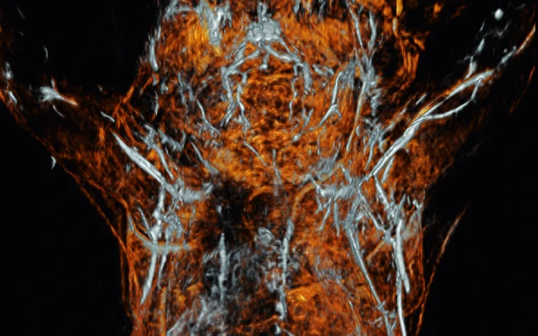
A European research team has published results on PAM3, an integrated 3D photoacoustic and ultrasound breast imaging system, in the journal Photoacoustics. The study, featured on the cover of Volume 50, details how the system combines light and sound to improve non-invasive imaging in dense breast tissue.
The paper, titled “Hybrid three-dimensional full-view multi-wavelength photoacoustic and ultrasound breast tomography”, presents a dual-modality system designed to solve a common problem in photoacoustic imaging: sound travelling at different speeds through different types of biological tissue, which distorts the final image.
How the Technology Works
Photoacoustic imaging works by combining light absorption with ultrasound detection. Short pulses of laser light illuminate the tissue, where natural absorbers, mainly hemoglobin in blood vessels, absorb the energy and expand slightly. This expansion creates weak ultrasound waves that travel back to the surface, where acoustic sensors detect them to build a 3D image of blood vessel networks.
Because tumors need new blood vessels (angiogenesis) to grow, mapping blood vessel density and blood oxygen levels gives doctors useful diagnostic details without using ionizing radiation or contrast agents.
However, breast tissue is a mix of fat, glandular tissue, and lesions, and sound travels through each at slightly different speeds. If an imaging system assumes a single average sound speed, the reconstructed image becomes blurry. The PAM3 system solves this by first using ultrasound to measure the exact sound speed at every point in the breast. It then uses this 3D sound map to correct the photoacoustic image, keeping blood vessel networks sharp and accurate.
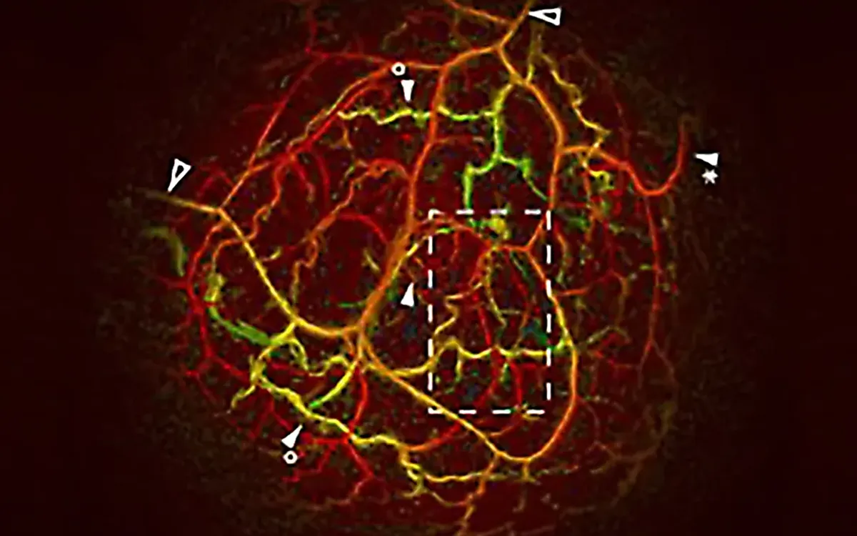
In vivo breast image made with the PAM3 system.
Optical Excitation: EKSPLA PhotoSonus Laser Integration
To deliver the required multi-wavelength optical pulses, the PAM3 setup incorporated an EKSPLA PhotoSonus series tunable wavelength laser as its photoacoustic excitation source. The source comprises two identical units, each consisting of a Q-switched Nd:YAG pump laser, a DKDP second harmonic generator, and a BBO optical parametric oscillator producing 5 ns pulses; their outputs are combined into a single beam coupled to the fiber bundle.
Designed specifically for biomedical and photoacoustic imaging applications, the PhotoSonus series provides a combined output energy from 450 mJ at 680 nm to 230 mJ at 1060 nm, roughly three times the energy reported for lasers in the same class, and 1.5 times per laser-OPO unit. Its fully motorized and computer-controlled architecture allowed for straightforward hardware integration into the PAM3 control software. Operational features used during the study included synchronized external trigger inputs and outputs, internal energy monitoring, a motorized optical attenuator, and automated switching between OPO signal and idler wavelengths.
Performance Results Reported in the Study
- Spatial Resolution: ~0.7 mm isotropic in 3D (786 µm in x, 775 µm in y, 693 µm in z), improving to 426 µm in z with 100 reconstruction iterations, comparable to the in-plane resolution of clinical breast MRI.
- Imaging Depth: Captures small blood vessels up to 48 mm deep inside tissue.
- Dual Data: Provides two complementary views at once: blood vessel distribution (optical) and tissue sound speed (ultrasound).
The project was carried out by a multi-institutional European collaboration of academic, industrial, and clinical partners, including the University of Twente (TechMed Centre), Center for Physical Sciences and Technology (FTMC), Centrum Wiskunde & Informatica (CWI), University College London (UCL), Brno University of Technology, P.A. Imaging R&D B.V., Imasonic SAS, Leiden University Medical Center (LUMC), Ziekenhuisgroep Twente (ZGT), and Medisch Spectrum Twente (MST).