PhotoSonus T
High Energy Table-Top Tunable Wavelength Lasers for Photoacoustic Imaging
PhotoSonus T is desktop version of high energy tunable laser source for photo-acoustic imaging. It features high output energies for imaging larger volumes of tissue.

Features
- Hands-free, automated wavelength tuning from 330 to 2600 nm
- Ultra-wide OPO signal tuning range from 660 to 1320 nm
- Up to 230 mJ in range 660 – 2600 nm,
35 mJ in range 330 – 660 nm - Narrow linewidth across tuning range
- 3 – 5 ns pulse duration
- Remote control via key pad or PC
- Separate output port for 532 nm beam. Output for 1064 nm is optional
- OPO pump energy monitoring
- Fast wavelength switching within entire signal or idler ranges
Applications
- Photoacoustic imaging
- Flash photolysis
- Photobiology
- Remote sensing
- Non-linear spectroscopy
Description
PhotoSonus T series tunable laser seamlessly integrates in a compact housing a nanosecond optical parametric oscillator and Nd:YAG Q-switched laser.
Three models with different output pulse energy values and different repetition rates are offered. The most powerful model has more than 230 mJ pulse energy. Narrow linewidth (<10 cm⁻¹) is nearly constant trough almost whole tuning range, which makes laser suitable for many spectroscopy application.
The device is controlled from the remote keypad or PC using LabVIEW™ drivers that are supplied with the system. The remote pad features a backlit display that is easy to read even while wearing laser safety glasses.
System is designed for easy and cost-effective maintenance. Replacement of flashlamps can be done without misalignment of the laser cavity and deterioration of laser performance. OPO pump energy monitoring system helps to increase lifetime of the optical components.
Benefits
- High pulse energy (up to 230 mJ) is highly beneficial for photoacoustics imaging applications
- Superior tuning resolution (1 – 2 cm⁻¹) allows recording of high quality spectra
- High integration level saves space in the laboratory
- Flashlamps replacement without misalignment of the laser cavity saves on maintenance costs
- In-house design and manufacturing of complete systems, including pump lasers, guarantees on-time warranty and post warranty services and spares supply
- Variety of control interfaces: USB, RS232, optional LAN and WLAN ensures easy control and integration with other equipment
- Attenuator and fiber bundle coupling options facilitate incorporation of PhotoSonus T systems into various experimental environments
Specifications
| Model | PhotoSonus T-10 | PhotoSonus T-20 | PhotoSonus T+ |
|---|---|---|---|
| OPO 1) | |||
| Wavelength range | |||
| Signal | 660 – 1320 nm | 660 – 1320 nm | 660 – 1064 nm 2) |
| Idler | 1065 – 2600 nm | 1065 – 2600 nm | 1065 – 2600 nm |
| SH (optional) | 330 – 660 nm | 330 – 660 nm | 330 – 530 nm (330 – 659 nm) 3) |
| Output max pulse energy 4) | |||
| OPO | 150 mJ | 130 mJ | 230 mJ |
| SH | 25 mJ | 21 mJ | 35 mJ |
| Linewidth 5) | < 15 cm‑1 | < 15 cm‑1 | < 15 cm‑1 |
| Tuning resolution 6) | |||
| Signal | 1 cm‑1 | 1 cm‑1 | 1 cm‑1 |
| Idler | 1 cm‑1 | 1 cm‑1 | 1 cm‑1 |
| SH | 2 cm‑1 | 2 cm‑1 | 2 cm‑1 |
| Pulse duration 7) | 3 – 5 ns | 3 – 5 ns | 3 – 5 ns |
| Typical beam diameter 8) | 7 mm | 7 mm | 9 mm |
| Typical beam divergence 9) | < 2 mrad | < 2 mrad | < 2 mrad |
| Polarization | |||
| Signal beam | horizontal | horizontal | horizontal |
| Idler beam | vertical | vertical | vertical |
| SH beam | vertical | vertical | vertical |
| Pump laser 10) | |||
| Pump wavelength | 532 nm | 532 nm | 532 nm |
| Pulse duration | 4 – 6 ns | 4 – 6 ns | 4 – 6 ns |
| Beam quality | ”Hat-Top” in near field. Close to Gaussian in far field | ”Hat-Top” in near field. Close to Gaussian in far field | ”Hat-Top” in near field. Close to Gaussian in far field |
| Beam divergence | < 0.6 mrad | < 0.6 mrad | < 0.6 mrad |
| Pulse energy stability (StdDev) | < 2.5 % | < 2.5 % | < 2.5 % |
| Pulse repetition rate | 10 Hz | 20 Hz | 10 Hz |
| Physical characteristics | |||
| Unit size (W × L × H mm) | 456 × 821 × 270 mm | 456 × 821 × 270 mm | 456 × 821 × 270 mm |
| Power supply size (W × L × H) | 330 × 490 × 585 mm | 330 × 490 × 585 mm | 330 × 490 × 585 mm |
| Umbilical length | 2.5 m | 2.5 m | 2.5 m |
| Operating requirements | |||
| Water consumption (max 20 °C) 11) | < 10 l/min | < 10 l/min | < 10 l/min |
| Room temperature | 18 – 27 °C | 18 – 27 °C | 18 – 27 °C |
| Relative humidity | 20 – 80 % (non-condensing) | 20 – 80 % (non-condensing) | 20 – 80 % (non-condensing) |
| Power requirements 8) | 200 – 240 VAC, single phase, 50/60 Hz | 200 – 240 VAC, single phase, 50/60 Hz | 200 – 240 VAC, single phase, 50/60 Hz |
| Power consumption | < 1.5 kVA | < 2.5 kVA | < 2.5 kVA |
| Cleanliness of the room | not worse than ISO Class 9 | not worse than ISO Class 9 | not worse than ISO Class 9 |
| Model | PhotoSonus T-10 | PhotoSonus T-20 | PhotoSonus T+ |
|---|
- Due to continuous improvement, all specifications are subject to change without notice. The parameters marked typical are not specifications. They are indications of typical performance and will vary with each unit we manufacture. Unless stated otherwise all specifications are measured at 700 nm and for basic system without options.
- Optional signal extended range: 660 – 1320 nm.
- When extended signal range is selected.
- See tuning curves for typical outputs at different wavelengths.
- At 700 nm or higher wavelengths.
- When wavelength is controlled from PC. When wavelength is controlled from keypad, tuning resolution is 0.1 nm for signal,
- 1 nm for idler and 0.5 nm for SH.
- FWHM measured with photodiode featuring 1 ns rise time and 300 MHz bandwidth oscilloscope.
- Beam diameter is measured at 700 nm at the 1/e2 level and can vary depending on the pump pulse energy.
- Full angle measured at the FWHM level at 700 nm.
- Separate output port for the 532 nm beam is standard. Output for 1064 nm beam is optional. Pump laser output will be optimized for the best OPO operation and specification may vary with each unit we manufacture.
- Air cooled power supply is available as option.
- Mains voltage should be specified when ordering.
Note: Laser must be connected to the mains electricity all the time. If there will be no mains electricity for longer that 1 hour then laser (system) needs warm up for a few hours before switching on.
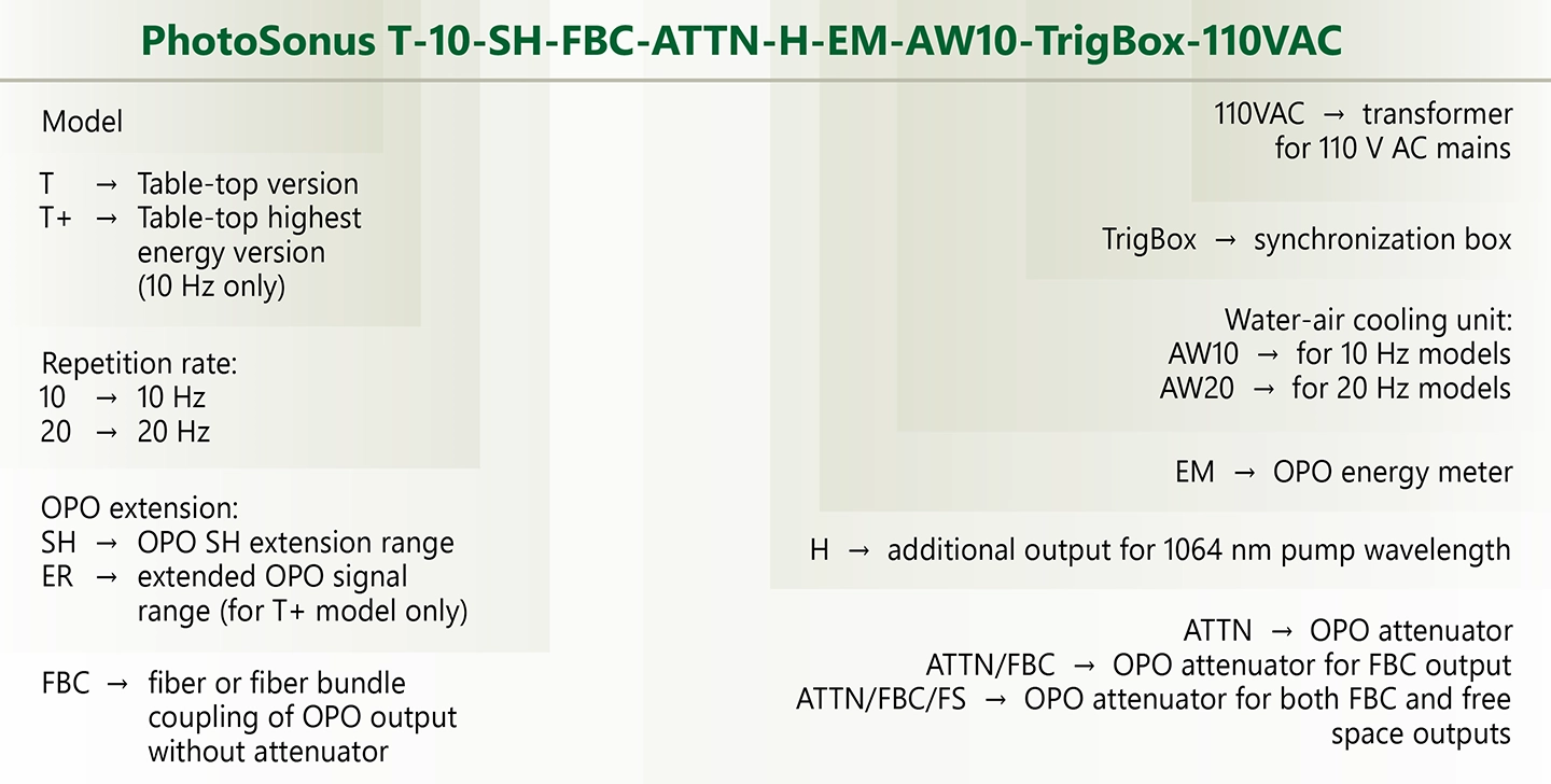
Ordering information of PhotoSonus T.
Options
| Option | Features |
|---|---|
| -SH | Efficient second harmonic generator for 330 – 660 nm range. |
| -ER | Extended OPO signal range (for T+ model only). |
| -FBC | Fiber bundle coupled output. |
| -ATTN | Pulse energy attenuator. |
| -H | Additional output for 1064 nm pump wavelength. |
| -EM | OPO energy meter. |
| -AW | Water-air cooled power supply. |
Performance
Publications
Breast cancer screening with mammography is less effective in women with dense breast tissue, prompting the use of ultrasound (US) imaging. While two- (2D) and three-dimensional (3D) US improve cancer detection, their low specificity leads to frequent unnecessary biopsies. Operator dependence on 2D US has led to the development of 3D automated breast volume scanners (ABVS), but challenges remain in distinguishing benign from malignant lesions. We developed a 3D photoacoustic and ultrasound (PAUS)–ABVS system that integrates a large field-of-view, 768-element transducer to improve diagnostic accuracy. In a clinical study of 61 patients with 36 benign and 30 malignant lesions, multispectral photoacoustic imaging was used to measure blood volume and oxygen saturation within lesions. When combined with standard US BI-RADS (breast imaging reporting and data system) scores, the system achieved a sensitivity of 96.7% and specificity of 66.7%. This performance matched the best outcomes of 2D PAUS and outperformed conventional US. Our results suggest that the PAUS-ABVS can support more accurate breast cancer diagnosis while reducing unnecessary biopsies.
Image-guided phototheranostics, including photothermal therapy and photoacoustic imaging using plasmonic nanoparticles, has attracted attention due to its remarkable photothermal conversion effects. In particular, considering the several benefits of biocompatibility, unique plasmon resonance, and tunable optical properties, gold nanoparticles of various shapes have been widely utilized as phototheranostic agents. However, applications in the near-infrared window have been limited due to the tendency of anisotropic gold nanoparticles to transform into spherical shapes under repetitive laser irradiation, which cause a shift in the localized surface plasmon resonance peak and reduces photothermal stability. To address these limitations, we introduce an Fe layer between Au nanodiscs to synthesize Au/Fe/Au trilayer uniform structured nanodiscs (AuFeAuNDs) using nanoimprint lithography. This approach aims to improve photostability and endow a magnetic targeting ability, enabling more accurate spatiotemporal regulation. The AuFeAuNDs primarily function as photothermal therapy agents and also serve as chemodynamic agents via the Fenton reaction specifically within the tumor microenvironment, which induces ferroptosis. Moreover, the AuFeAuNDs trigger immunogenic cell death following photothermal therapy. This study demonstrates applications of magnetic-targeted AuFeAuNDs for photoacoustic imaging-guided, spatiotemporal-controlled photothermal therapy, chemodynamic therapy, and immune modulation to bolster anti-tumor immune responses in cancer treatment.
Fabry–Perot (FP) ultrasound sensors are a class of optical ultrasound sensors used in photoacoustic tomography (PAT) and other applications. Conventionally, an FP ultrasound sensor comprises an ultrasonically compressible planar microcavity, locally interrogated by a focused Gaussian beam. One way to increase the sensitivity could be to replace this beam with a Bessel beam. The rationale is twofold. First, as a Bessel beam’s wavefront better matches the modes of the planar microcavity, this could increase the -factor, leading to higher sensitivity. Second, as a Bessel beam provides a focused spatial structure—a central core surrounded by concentric rings—it might retain the ability to locally interrogate the sensor. To explore this idea, we developed an experimental system featuring a custom FP ultrasound sensor interrogated by a Bessel beam and evaluated its on-axis sensitivity, directivity, and image resolution when performing PAT. For comparison, we measured the same characteristics using a conventional focused Gaussian beam with a spot size similar to the core of the Bessel beam. As anticipated, the Bessel beam provided a higher ultrasonic on-axis sensitivity. However, the directivity and spatial resolution were degraded, suggesting that the Bessel beam yielded a larger acoustic element. We conclude that it is feasible to increase the sensitivity of an FP ultrasound sensor using a Bessel beam. Further work is required to establish whether differently designed Bessel beams could concurrently offer a smaller acoustic element.
Photoacoustic tomography (PAT) has the potential to become a widely used imaging tool in preclinical studies of small animals. This is because it can provide non-invasive, label free images of whole-body mouse anatomy, in a manner which is challenging for more established imaging modalities. However, existing PAT scanners are limited because they either do not implement a full 3-D tomographic reconstruction using all the recorded photoacoustic (PA) data and/or do not record the available 3-D PA time-series data around the mouse with sufficiently high spatial resolution ( ∼100μ m), which compromises image quality in terms of resolution, imaging depth and the introduction of artefacts. In this study, we address these limitations by demonstrating an all-optical, multi-view Fabry-Perot based scanner for whole body small animal imaging. The scanner densely samples the acoustic field with a large number of detection points (>100,000), evenly distributed around the mouse. The locations of the detection points were registered onto a common coordinate system, before a tomographic reconstruction using all the recorded PA time series was implemented. This enabled the acquisition of high resolution, whole-body PAT images of ex-vivo mice, with anatomical features visible across the entire cross section.
Photoacoustic imaging offers high spatial resolution, imaging depth, and molecular information, emerging as a promising biomedical imaging modality. In particular, when using exogenous contrast, the advantages of photoacoustic imaging can be more effectively utilized in preclinical and clinical studies. We provide a novel approach to screen and identify efficient photoacoustic performance as contrast agents of the metals. To accomplish this, we introduce a novel figure of merit that quantifies the potential performance of contrast agents. As a result of the quantification, we discover that Ti nanodiscs outperform Pt nanodiscs in terms of photoacoustic ability, which shows a similar level to Au nanodiscs. We compare these results by performing a photoacoustic phantom imaging experiment. The photoacoustic performance of the three materials is compared by comparing the signal intensity of the materials measured on the photoacoustic image for various wavelengths. The imaging results further support our findings, demonstrating the superior performance of Ti nanodiscs as contrast agents.
Photonic-based methods are crucial in biology and medicine due to their non-invasive nature, allowing remote measurements without affecting biological specimens. The study of diatoms using advanced photonic methods remains a relatively underexplored area, presenting significant opportunities for pioneering discoveries. This research provides a comprehensive analysis of marine diatoms, specifically Nitzschia sp., across varying salinity levels, integrating fluorescence lifetime imaging microscopy (FLIM), combined photoacoustic and fluorescence tomographies (PAFT), and ultrastructural examinations using transmission electron microscopy. Key findings include a systematic shift in the mean fluorescence lifetime from 570 ps at 20‰ to 940 ps at 80‰, indicating functional adaptations in chlorophyll molecules within light-harvesting complexes. At 60‰ salinity, anomalies are observed in the development of silica valves and polysaccharide layers, suggesting abnormalities in valve morphogenesis. Lipid droplets within the cells display a minimum diameter at 40‰, indicating metabolic adjustments to osmotic stress. The intensity of both fluorescence and photoacoustic signals increases with increasing salinity levels. These insights enhance understanding of the ecological implications of salinity stress on diatom communities and pave the way for future research on leveraging the unique adaptive mechanisms of microalgae for environmental monitoring and sustainable biotechnological applications.
Lewis hunting reaction refers to the alternating cold-induced vasoconstriction and dilation in extremities, whose underlying mechanism is complex. While numerous studies reported this intriguing phenomenon by measuring cutaneous temperature fluctuation under cold exposure, few of them tracked peripheral vascular responses in real-time, lacking a non-invasive and quantitative imaging tool. To better monitor hunting reaction and diagnose relevant diseases, we developed a hybrid photoacoustic ultrasound (PAUS) tomography system to monitor finger vessels’ dynamic response to cold, together with simultaneous temperature measurement. We also came out a standard workflow for image analysis with self-defined indices. In the small cohort observational study, vascular changes in the first cycle of hunting reaction were successfully captured by the image series and quantified. Time difference between vasodilation and temperature recovery was noticed and reported for the first time, thanks to the unique capability of the PAUS imaging system in real-time and continuous vascular monitoring. The developed imaging system and indices enabled more objective and quantitative monitoring of peripheral vascular activities, indicating its great potential in numerous clinical applications.
SignificanceEndocavity ultrasound (US) imaging is a frequently employed diagnostic technique in gynecology and urology for the assessment of male and female genital diseases that present challenges for conventional transabdominal imaging. The integration of photoacoustic (PA) imaging with clinical US imaging has displayed promising outcomes in clinical research. Nonetheless, its application has been constrained due to size limitations, restricting it to spatially confined locations such as vaginal or rectal canals.AimThis study presents the development of a video-rate (20 Hz) endocavity PA/harmonic US imaging (EPAUSI) system.ApproachThe approach incorporates a commercially available endocavity US probe with a miniaturized laser delivery unit, comprised of a single large-core fiber and a line beamshaping engineered diffuser. The system facilitates real-time image display and subsequent processing, including angular energy density correction and spectral unmixing, in offline mode.ResultsThe spatial resolutions of the concurrently acquired PA and harmonic US images were measured at 318 μm and 291 μm in the radial direction, respectively, and 1.22 deg and 1.50 deg in the angular direction, respectively. Furthermore, the system demonstrated its capability in multispectral PA imaging by successfully distinguishing two clinical dyes in a tissue-mimicking phantom. Its rapid temporal resolution enabled the capture of kinetic dye perfusion into an ex vivo porcine ovary through the depth of porcine uterine tissue. EPAUSI proved its clinical viability by detecting pulsating hemodynamics in the male rat’s prostate in vivo and accurately classifying human blood vessels into arteries and veins based on sO2 measurements.ConclusionsOur proposed EPAUSI system holds the potential to unveil previously overlooked indicators of vascular alterations in genital cancers or endometriosis, addressing pressing requirements in the fields of gynecology and urology.
Photoacoustic tomography (PAT) is increasingly being used for high-resolution biological imaging at depth. Signal-to-noise ratios and resolution are the main factors that determine image quality. Various reconstruction algorithms have been proposed and applied to reduce noise and enhance resolution, but the efficacy of signal preprocessing methods which also affect image quality, are seldom discussed. We, therefore, compared common preprocessing techniques, namely bandpass filters, wavelet denoising, empirical mode decomposition, and singular value decomposition. Each was compared with and without accounting for sensor directivity. The denoising performance was evaluated with the contrast-to-noise ratio (CNR), and the resolution was calculated as the full width at half maximum (FWHM) in both the lateral and axial directions. In the phantom experiment, counting in directivity was found to significantly reduce noise, outperforming other methods. Irrespective of directivity, the best performing methods for denoising were bandpass, unfiltered, SVD, wavelet, and EMD, in that order. Only bandpass filtering consistently yielded improvements. Significant improvements in the lateral resolution were observed using directivity in two out of three acquisitions. This study investigated the advantages and disadvantages of different preprocessing methods and may help to determine better practices in PAT reconstruction.
Although spherical gold (Au) nanoparticles have remarkable photothermal conversion efficiency and photostability, their weak absorption in the near-infrared (NIR) region and poor penetration into deep tissues have limited further applications to NIR light-mediated photoacoustic (PA) imaging and noninvasive photothermal cancer therapy. Here, we developed bimetallic hyaluronate-modified Au–platinum (HA-Au@Pt) nanoparticles for noninvasive cancer theranostics by NIR light-mediated PA imaging and photothermal therapy (PTT). The growth of Pt nanodots on the surface of spherical Au nanoparticles enhanced the absorbance in the NIR region and broadened the absorption bandwidth of HA-Au@Pt nanoparticles by the surface plasmon resonance (SPR) coupling effect. In addition, HA facilitated the transdermal delivery of HA-Au@Pt nanoparticles through the skin barrier and enabled clear tumor-targeted PA imaging. Compared to conventional PTT via injection, HA-Au@Pt nanoparticles were noninvasively delivered into deep tumor tissues and completely ablated the targeted tumor tissues by NIR light irradiation. Taken together, we could confirm the feasibility of HA-Au@Pt nanoparticles as a NIR light-mediated biophotonic agent for noninvasive skin cancer theranostics.