Mobile, Tunable Wavelength Laser Source for Photoacoustic Imaging PhotoSonus M
High Energy, Mobile, Tunable Wavelength Laser Source for Photoacoustic Imaging
Following the demand for high output energies in the photoacoustic market for imaging larger volumes of tissue, PhotoSonus M, an updated high energy tunable laser source for photo-acoustic imaging, was introduced.

Features
- High up to 250 mJ output energy
- Wide tuning range from 330 to 2300 nm
- Ultra-wide OPO signal tuning range from 660 to 1320 nm
- 10 Hz pulse repetition rate
- Integrated pump laser, OPO and PSU in single mobile unit
- Low maintenance cost
- Fiber bundle connectors with safety interlock
- Fast wavelength switching within entire signal or idler range between two consecutive pulses
- Integrated energy meter (optional)
- Motorized attenuator (optional)
- Access to pump laser wavelengths 1064/532 nm (optional)

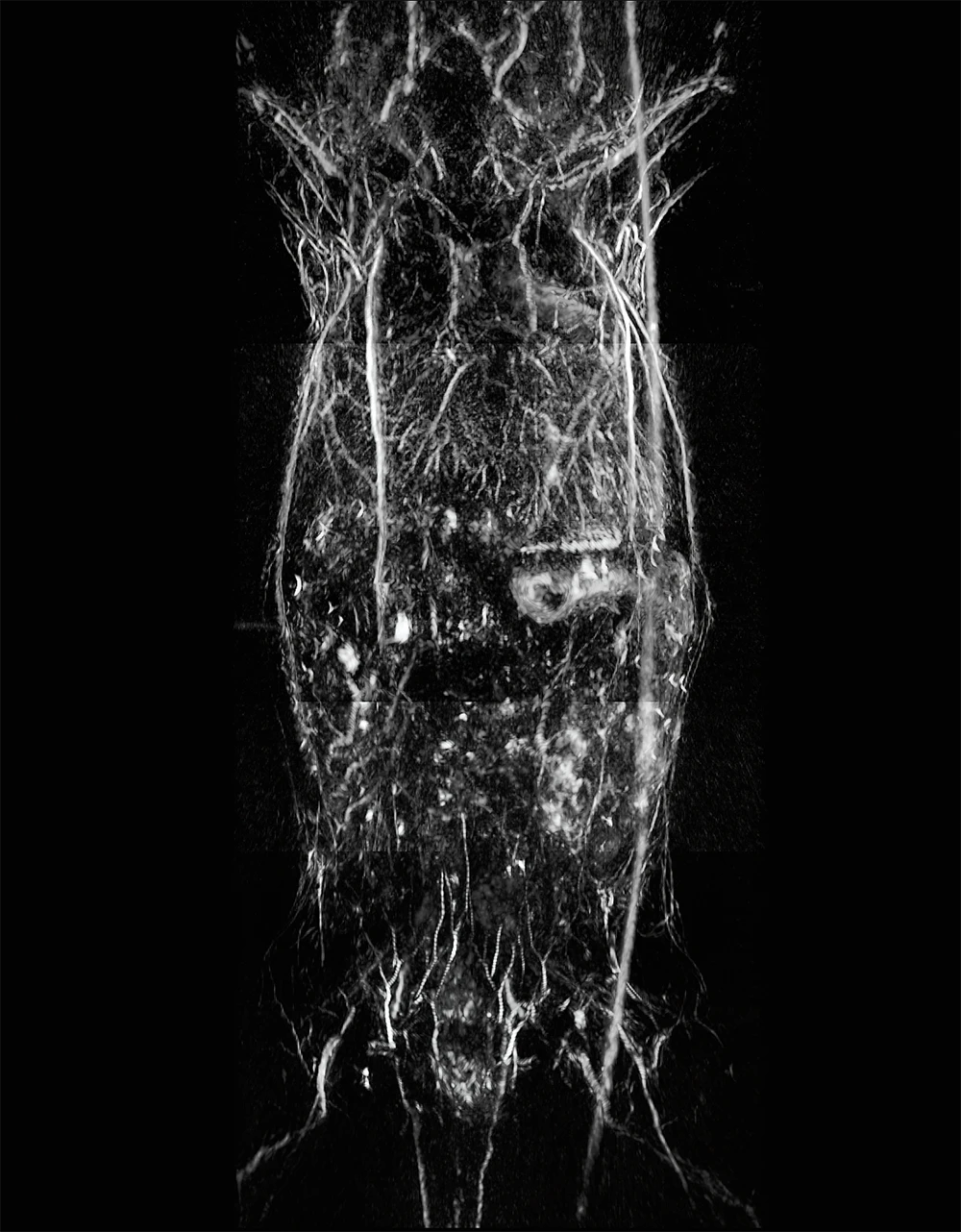
Photoacoustic image of the upper torso and brain of a female mouse.
Courtesy of PhotoSound Technologies, Inc.
Photoacoustic image of a mouse.
Courtesy of PhotoSound Technologies, Inc.
Description
Following the demand for high output energies in the photoacoustic market for imaging larger volumes of tissue, PhotoSonus M, an updated high energy tunable laser source for photo-acoustic imaging, was introduced.
Time-tested EKSPLA nanosecond pump laser, parametric oscillator, power supply and cooling unit are integrated in a single robust housing to provide mobility, ease of use and low maintenance cost. The highly flexible PhotoSonus M platform makes it easily integrated and used in a photoacoustic imaging system. It is fully motorized and computer controlled, with user trigger outputs and inputs and special options such as motorized switching between OPO signal and idler, motorized attenuator, internal energy meter and electromechanical output shutter.
Recently, a fast wavelength switching option was introduced that enables each laser pulse to have a different wavelength within the entire signal or idler range and at any sequence. This new feature, combining high pulse energy (up to 180 mJ) and wide wavelength tuning range (330 – 2300 nm) makes PhotoSonus M the irreplaceable imaging source for any photo acoustic system.
For even higher sample imaging depth and resolution a PhotoSonus M+, with up to 250 mJ maximum pulse energy, was introduced.
For convenience, the outputs of PhotoSonus M and PhotoSonus M+ lasers can be coupled with almost any type of fiber bundle.
| Model | PhotoSonus M-10 | PhotoSonus M+ |
|---|---|---|
| OPO 1) | ||
| Wavelength range | ||
| Signal | 660 – 1320 nm | 660 – 1064 nm 2) |
| SH extension range (optional) 3) | 330 – 659 nm | 330 – 530 nm (330 – 659 nm 4) ) |
| Idler (optional) | 1065 – 2300 nm | 1065 – 2300 nm |
| OPO output MAX pulse energy 5) | > 180 mJ | > 250 mJ |
| Pulse repetition rate | 10 Hz | 10 Hz |
| Scanning step | ||
| Signal | 0.1 nm | 0.1 nm |
| Idler | 1 nm | 1 nm |
| Pulse duration 6) | 3 – 5 ns | 3 – 5 ns |
| Signal linewidth 7) | < 15 cm‑1 | < 15 cm‑1 |
| Typical signal beam diameter (1/e2) 8) | 7 ± 2 mm | 9 ± 2 mm |
| Physical characteristics | ||
| Unit size (W × L × H mm) | 434 × 672 × 887 mm | 434 × 672 × 887 mm |
| Operating requirements | ||
| Room temperature | 18 – 27 °C | 18 – 27 °C |
| Relative humidity | 20 – 80 % (non-condensing) | 20 – 80 % (non-condensing) |
| Power requirements 9) | 200 – 240 VAC, single phase, 50/60 Hz | 200 – 240 VAC, single phase, 50/60 Hz |
| Power consumption | < 1.5 kVA | < 2.5 kVA |
| Model | PhotoSonus M-10 | PhotoSonus M+ |
|---|
- Due to continuous improvement, all specifications are subject to change without notice. The parameters marked typical are not specifications. They are indications of typical performance and will vary with each unit we manufacture. Unless stated otherwise all specifications are measured at 700 nm.
- Optional signal extended range: 660 – 1320 nm.
- This option results lower pulse energy in the main OPO range. Please contact EKSPLA for more details.
- When extended signal range is selected.
- Measured at the free space output. See tuning curves for typical energy levels at different wavelengths.
- FWHM measured with photodiode featuring 1 ns rise time and 300 MHz bandwidth oscilloscope.
- At 700 nm or higher wavelengths.
- Measured at the free space output at 700 nm. Can be adjusted as per request.
- Mains voltage should be specified when ordering.
Note: Laser must be connected to the mains electricity all the time. If there will be no mains electricity for longer that 1 hour then laser (system) needs warm up for a few hours before switching on.
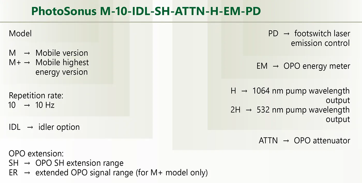
Ordering information of PhotoSonus M laser sources.
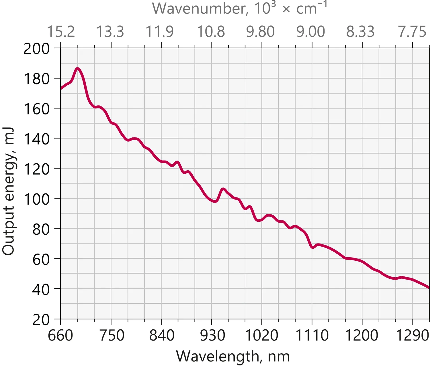
Typical PhotoSonus M-10 extended signal output pulse energy vs. wavelength curve.
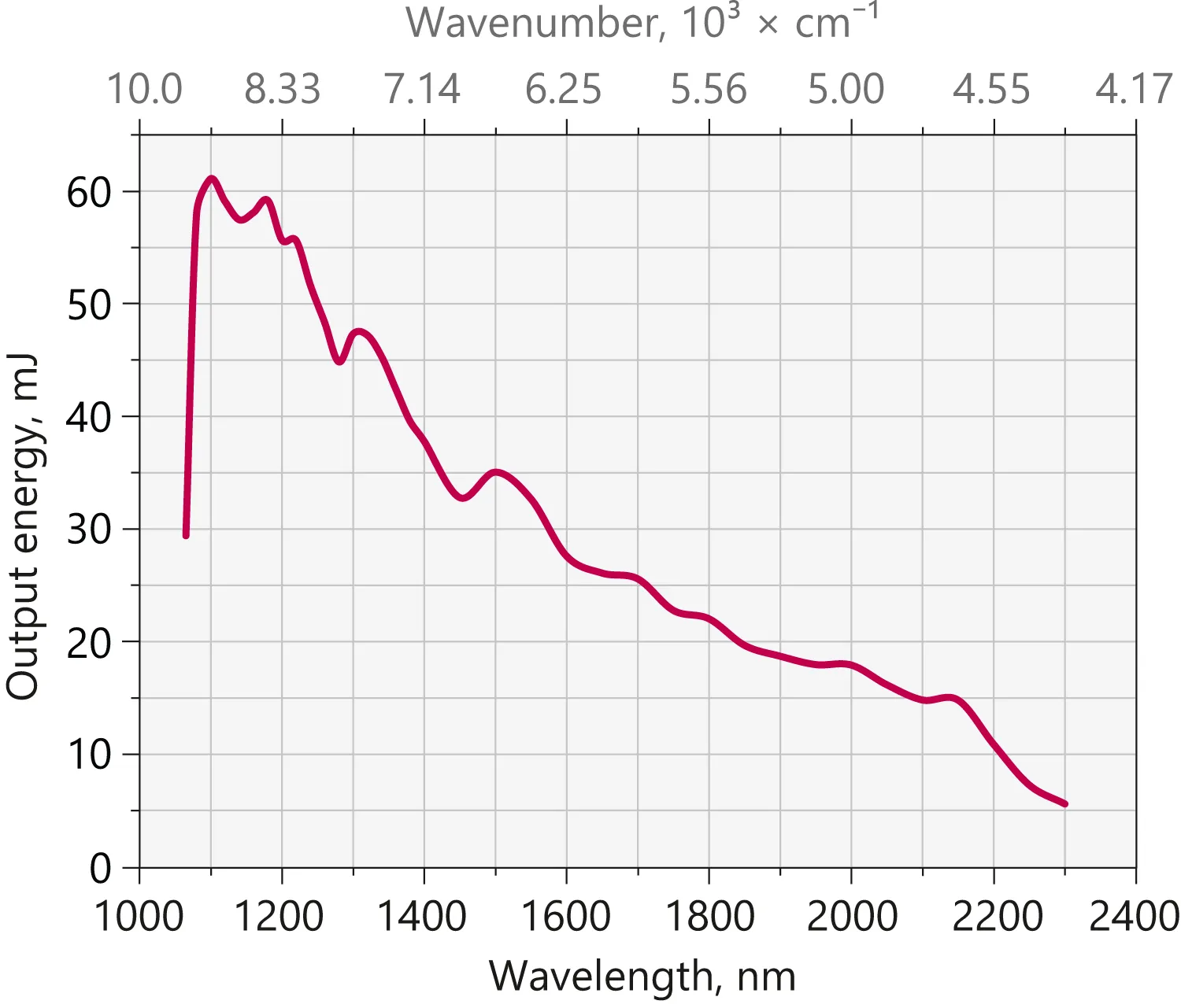
Typical PhotoSonus M-10 idler output pulse energy vs. wavelength curve,
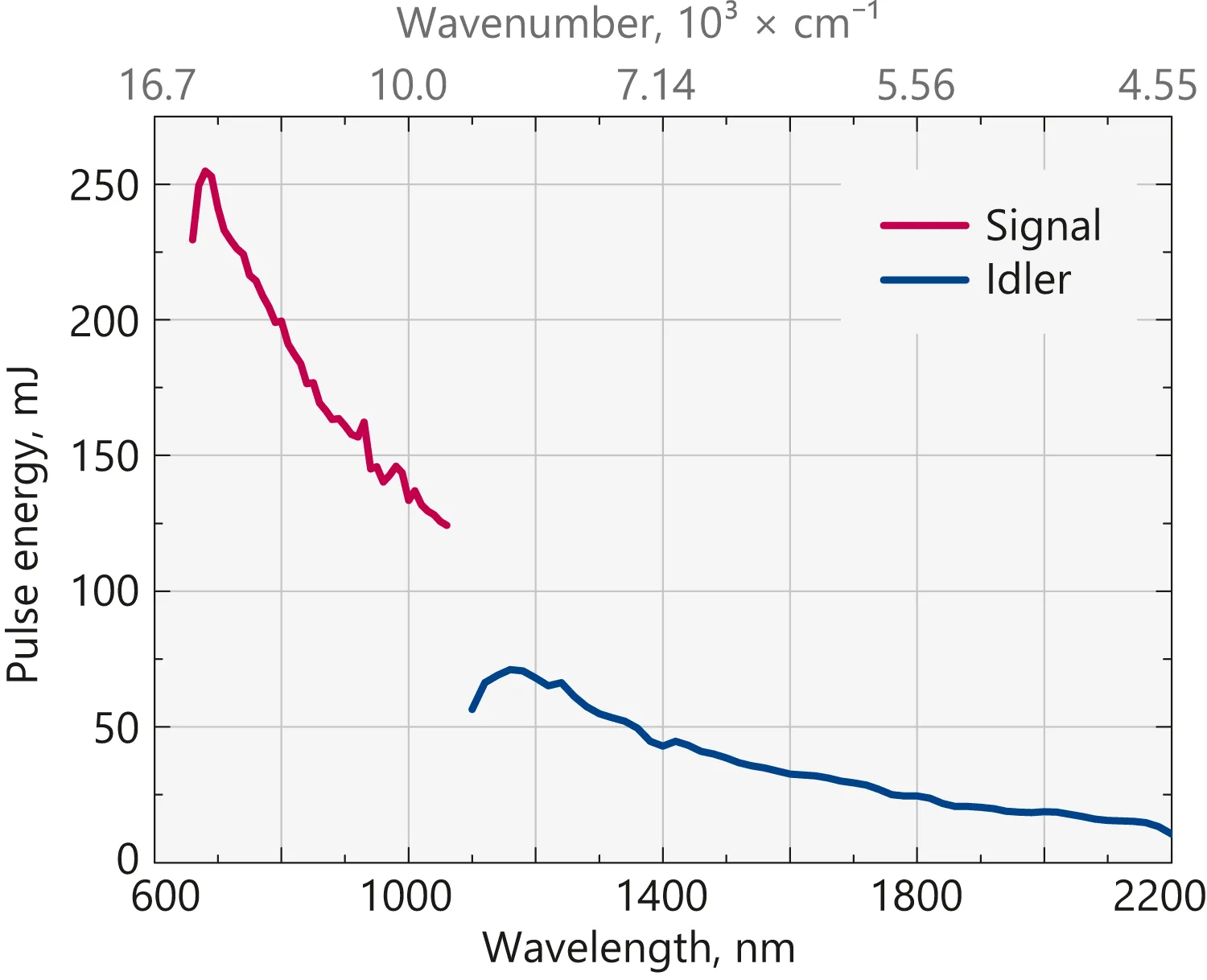
Typical PhotoSonus M+ signal and idler output pulse energy vs. wavelength curve.

PhotoSonus M outline drawings.
All dimensions are in millimetres.