Imaging with laser-induced ultrasound
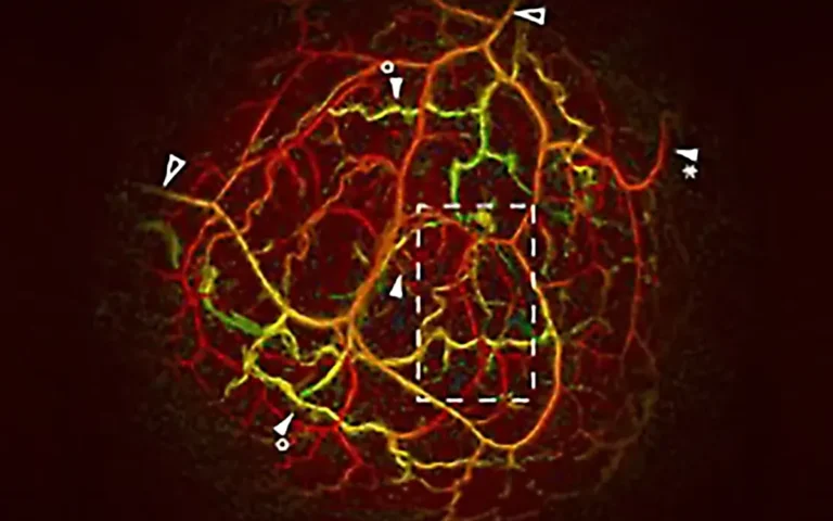
Photoacoustic imaging reads optical absorption at ultrasonic depth. The tissue is illuminated with tunable-wavelength laser pulses; where the light is absorbed, it heats slightly, expands thermoelastically and emits an ultrasound wave. Ultrasonic transducers detect these waves, and 2D or 3D images are reconstructed from the accumulated data.
The result is high-contrast, non-invasive imaging of living tissue, used in pre-clinical research and clinical practice alike. Laser sources for photoacoustic imaging include lamp-pumped and diode-pumped solid-state (DPSS) tunable-wavelength OPO systems with pulse energies up to 250 mJ.

Photoacoustic image of the upper torso and brain of a female mouse.
Courtesy of PhotoSound Technologies, Inc.

Photoacoustic image of the mouse liver.
Courtesy of PhotoSound Technologies, Inc.