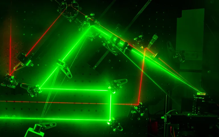
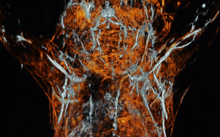
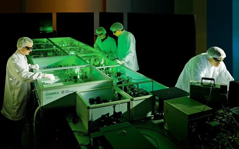
Photoacoustic imaging is a valuable high-contrast in vivo imaging technique for pre-clinical and clinical applications. This technique uses laser-induced ultrasound.
Ultrasound signal is generated in tissue, when it absorbs tunable wavelength laser light and expands thermo-elastically, and their waves are detected by ultrasonic transducers. 2D or 3D images are then reconstructed from the accumulated data.
Laser sources for photoacoustic imaging include lamp-pumped tunable wavelength and diode-pumped solid-state (DPSS) tunable wavelength OPO systems.
Publications
Wide-field three-dimensional photoacoustic/ultrasound scanner using a two-dimensional matrix transducer array
Related products: PhotoSonus M PhotoSonus X
Two-dimensional matrix transducer arrays are the most appropriate imaging probes for acquiring dual-modal 3D photoacoustic (PA)/ultrasound (US) images. However, they have small footprints which limit the field-of-view (FOV) to less than 10 mm × 10 mm and degrade the spatial resolution. In this study, we demonstrate a dual-modal PA and US imaging system (using a 2D matrix transducer array and a motorized 2D scanning system) to enlarge the FOV of volumetric images. Multiple PA volumes were merged to form a wide-field image of approximately 45 mm × 45 mm. In vivo imaging was demonstrated using rat sentinel lymph nodes (SLNs) and bladders stained with methylene blue. We believe that this volumetric PA/US imaging technique with a 2D matrix transducer array can be a useful tool for narrow-field real-time monitoring and wide-field imaging of various preclinical and clinical studies.
An Investigation of Signal Preprocessing for Photoacoustic Tomography
Related products: PhotoSonus M PhotoSonus X
Photoacoustic tomography (PAT) is increasingly being used for high-resolution biological imaging at depth. Signal-to-noise ratios and resolution are the main factors that determine image quality. Various reconstruction algorithms have been proposed and applied to reduce noise and enhance resolution, but the efficacy of signal preprocessing methods which also affect image quality, are seldom discussed. We, therefore, compared common preprocessing techniques, namely bandpass filters, wavelet denoising, empirical mode decomposition, and singular value decomposition. Each was compared with and without accounting for sensor directivity. The denoising performance was evaluated with the contrast-to-noise ratio (CNR), and the resolution was calculated as the full width at half maximum (FWHM) in both the lateral and axial directions. In the phantom experiment, counting in directivity was found to significantly reduce noise, outperforming other methods. Irrespective of directivity, the best performing methods for denoising were bandpass, unfiltered, SVD, wavelet, and EMD, in that order. Only bandpass filtering consistently yielded improvements. Significant improvements in the lateral resolution were observed using directivity in two out of three acquisitions. This study investigated the advantages and disadvantages of different preprocessing methods and may help to determine better practices in PAT reconstruction.
Size-tunable ICG-based contrast agent platform for targeted near-infrared photoacoustic imaging
Related products: PhotoSonus M PhotoSonus X
Near-infrared photoacoustic imaging (NIR-PAI) combines the advantages of optical and ultrasound imaging to provide anatomical and functional information of tissues with high resolution. Although NIR-PAI is promising, its widespread use is hindered by the limited availability of NIR contrast agents. J-aggregates (JA) made of indocyanine green dye (ICG) represents an attractive class of biocompatible contrast agents for PAI. Here, we present a facile synthesis method that combines ICG and ICG-azide dyes for producing contrast agents with tunable size down to 230 nm and direct functionalization with targeting moieties. The ICG-JA platform has a detectable PA signal in vitro that is two times stronger than whole blood and high photostability. The targeting ability of ICG-JA was measured in vitro using HeLa cells. The ICG-JA platform was then injected into mice and in vivo NIR-PAI showed enhanced visualization of liver and spleen for 90 min post-injection with a contrast-to-noise ratio of 2.42.
Deep Learning Enhances Multiparametric Dynamic Volumetric Photoacoustic Computed Tomography In Vivo (DL-PACT)
Related products: PhotoSonus M PhotoSonus X
Photoacoustic computed tomography (PACT) has become a premier preclinical and clinical imaging modality. Although PACT's image quality can be dramatically improved with a large number of ultrasound (US) transducer elements and associated multiplexed data acquisition systems, the associated high system cost and/or slow temporal resolution are significant problems. Here, a deep learning-based approach is demonstrated that qualitatively and quantitively diminishes the limited-view artifacts that reduce image quality and improves the slow temporal resolution. This deep learning-enhanced multiparametric dynamic volumetric PACT approach, called DL-PACT, requires only a clustered subset of many US transducer elements on the conventional multiparametric PACT. Using DL-PACT, high-quality static structural and dynamic contrast-enhanced whole-body images as well as dynamic functional brain images of live animals and humans are successfully acquired, all in a relatively fast and cost-effective manner. It is believed that the strategy can significantly advance the use of PACT technology for preclinical and clinical applications such as neurology, cardiology, pharmacology, endocrinology, and oncology.
Low-intensity ultrasound is an emerging modality for neuromodulation. Yet, transcranial neuromodulation using low-frequency piezo-based transducers offers poor spatial confinement of excitation volume, often bigger than a few millimeters in diameter. In addition, the bulky size limits their implementation in a wearable setting and prevents integration with other experimental modalities. Here, we report spatially confined optoacoustic neural stimulation through a miniaturized Fiber-Optoacoustic Converter (FOC). The FOC has a diameter of 600 μm and generates omnidirectional ultrasound wave locally at the fiber tip through the optoacoustic effect. We show that the acoustic wave generated by FOC can directly activate individual cultured neurons and generate intracellular Ca2+ transients. The FOC activates neurons within a radius of 500 μm around the fiber tip, delivering superior spatial resolution over conventional piezo-based low-frequency transducers. Finally, we demonstrate direct and spatially confined neural stimulation of mouse brain and modulation of motor activity in vivo.
High-resolution multimodal photoacoustic microscopy and optical coherence tomography image-guided laser induced branch retinal vein occlusion in living rabbits
Related products: NT242 series
Joint high-resolution multimodal photoacoustic microscopy (PAM) and optical coherence tomography (OCT) was developed to improve the efficiency for visualizing newly developed retinal neovascularization (RNV) and to monitor the dynamic changes of retinal vein occlusion (RVO) in living rabbits. The RNV and RVO models were created in New Zealand rabbits by Rose Bengal laser-induced RVO. Dual modalities imaging equipment, including color fundus photography, fluorescein angiography (FA), OCT, and PAM, was used to image and assess the changes of retinal vasculature. In vivo experimental results exhibited that not only the treatment boundaries and the position of the occluded vasculature but also the structure of individual RNV were markedly observed using PAM platform with great resolution and high image contrast. The laser light energy of 80 nJ was used to induce photoacoustic signal, which is approximately half the energy of the American National Standards Institute safety limit. A cross-sectional structure of RNV was identified with the OCT modality. Furthermore, vibrant transformations in the RNV and the retinal morphology were examined at different times after laser occlusion: days 4, 28, 35, 49, and 90. PAM revealed high contrast and high resolution vascular imaging of the retina and choroid with amplified penetration depth. Through the present custom-built imaging system, both RNV and RVO can be reconstructed and observed in two and three dimensions. A unique dual modality A unique dual modality PAM and OCT can help precisely visualize and distinguish individual microvessels, microvessel depth, and the surrounding anatomy. Thus, the proposed multimodal ocular imaging platform may offer a potential equipment to enhance classification of microvasculature in a reliable and proficient manner in larger rabbit eyes.
Photoacoustic/Ultrasound/Optical Coherence Tomography Evaluation of Melanoma Lesion and Healthy Skin in a Swine Model
Related products: NL230 series
The marked increase in the incidence of melanoma coupled with the rapid drop in the survival rate after metastasis has promoted the investigation into improved diagnostic methods for melanoma. High-frequency ultrasound (US), optical coherence tomography (OCT), and photoacoustic imaging (PAI) are three potential modalities that can assist a dermatologist by providing extra information beyond dermoscopic features. In this study, we imaged a swine model with spontaneous melanoma using these modalities and compared the images with images of nearby healthy skin. Histology images were used for validation.
High-resolution, high-contrast mid-infrared imaging of fresh biological samples with ultraviolet-localized photoacoustic microscopy
Related products: NT242 series NT270 series
Mid-infrared (MIR) microscopy provides rich chemical and structural information about biological samples, without staining. Conventionally, the long MIR wavelength severely limits the lateral resolution owing to optical diffraction; moreover, the strong MIR absorption of water ubiquitous in fresh biological samples results in high background and low contrast. To overcome these limitations, we propose a method that employs photoacoustic detection highly localized with a pulsed ultraviolet laser on the basis of the Grüneisen relaxation effect. For cultured cells, our method achieves water-background suppressed MIR imaging of lipids and proteins at ultraviolet resolution, at least an order of magnitude finer than the MIR diffraction limits. Label-free histology using this method is also demonstrated in thick brain slices. Our approach provides convenient high-resolution and high-contrast MIR imaging, which can benefit the diagnosis of fresh biological samples.
Contrast Agent Enhanced Multimodal Photoacoustic Microscopy and Optical Coherence Tomography for Imaging of Rabbit Choroidal and Retinal Vessels in vivo
Related products: NT242 series
Multimodal imaging with photoacoustic microscopy (PAM) and optical coherence tomography (OCT) can be an effective method to evaluate the choroidal and retinal microvasculature. To improve the efficiency for visualizing capillaries, colloidal gold nanoparticles (AuNPs) have been applied as a multimodal contrast agent for both OCT and PAM imaging by taking advantage of the strong optical scattering and the strong optical absorption of AuNPs due to their surface plasmon resonance. Ultra-pure AuNPs were fabricated by femtosecond laser ablation, capped with polyethylene glycol (PEG), and administered to 13 New Zealand white rabbits and 3 Dutch Belted pigmented rabbits. The synthesized PEG-AuNPs (20.0 ± 1.5 nm) were demonstrated to be excellent contrast agents for PAM and OCT, and do not demonstrate cytotoxicity to bovine retinal endothelial cells in cell studies. The image signal from the retinal and choroidal vessels in living rabbits was enhanced by up to 82% for PAM and up to 45% for OCT, respectively, by the administered PEG-AuNPs, which enables detection of individual blood vessels by both imaging modalities. The biodistribution study demonstrated the AuNP accumulated primarily in the liver and spleen. Histology and TUNEL staining did not indicate cell injury or death in the lung, liver, kidney, spleen, heart, or eyes up to seven days after AuNP administration. PEG-AuNPs offer an efficient and safe contrast agent for multimodal ocular imaging to achieve improved characterization of microvasculature.
Photoacoustic transfection consists in the use of photoacoustic waves, generated in the thermoelastic expansion of a confined material absorbing a short pulse of a laser, to produce temporary mechanical deformations of the cell membrane and facilitate the delivery of plasmid DNA into cells. We show that high stress gradients, produced when picosecond laser pulses with a fluence of 100 mJ/cm2 are absorbed by piezophotonic materials, enable transfection of a plasmid DNA encoding Green Fluorescent Protein (gWizGFP, 3.74 MDa) in COS-7 monkey fibroblast cells with an efficiency of 5% at 20 °C, in 10 minutes. We did not observe significant cytotoxicity under these conditions. Photoacoustic transfection is scalable, affordable, enables nuclear localization and the dosage is easily controlled by the laser parameters.
High-resolution, in vivo multimodal photoacoustic microscopy, optical coherence tomography, and fluorescence microscopy imaging of rabbit retinal neovascularization
Related products: NT242 series
Photoacoustic microscopy (PAM) is an emerging imaging technology that can non-invasively visualize ocular structures in animal eyes. This report describes an integrated multimodality imaging system that combines PAM, optical coherence tomography (OCT), and fluorescence microscopy (FM) to evaluate angiogenesis in larger animal eyes. High-resolution in vivo imaging was performed in live rabbit eyes with vascular endothelial growth factor (VEGF)-induced retinal neovascularization (RNV). The results demonstrate that our multimodality imaging system can non-invasively visualize RNV in both albino and pigmented rabbits to determine retinal pathology using PAM and OCT and verify the leakage of neovascularization using FM and fluorescein dye. This work presents high-resolution visualization of angiogenesis in rabbits using a multimodality PAM, OCT, and FM system and may represent a major step toward the clinical translation of the technology.
Hydrophones based on interferometric fiber-optic sensors with applications in photoacoustics
Related products: NT350 series PhotoSonus M PhotoSonus X
Biomedical imaging used for medical diagnosis constantly requires improvement in the characteristics for imaging devices. The sensing devices are one of the most important pieces to improve in order to get images with better quality. In this thesis, it is proposed the use of interferometric fiber-optic sensors (which offer the advantages inherent to optical fibers) as devices to detect pressure/acoustic signals generated by the photoacoustic effect. It is explored the capability of using fiber-optic interferometric hydrophones in order to determine the thickness of a material derived from the acoustic signal generated when a sample is illuminated. In addition, the analysis of photoacoustic signals generated by the excitation of nanoparticles of an anisotropic material as absorption centers. Finally, the cross-section of a metallic sample was photoacoustically imaged by acquiring the pressure signals generated.
Photoacoustic imaging of voltage responses beyond the optical diffusion limit
Related products: NT242 series
Non-invasive optical imaging of neuronal voltage response signals in live brains is constrained in depth by the optical diffusion limit, which is due primarily to optical scattering by brain tissues. Although photoacoustic tomography breaks this limit by exciting the targets with diffused photons and detecting the resulting acoustic responses, it has not been demonstrated as a modality for imaging voltage responses. In this communication, we report the first demonstration of photoacoustic voltage response imaging in both in vitro HEK-293 cell cultures and in vivo mouse brain surfaces. Using spectroscopic photoacoustic tomography at isosbestic wavelengths, we can separate voltage response signals and hemodynamic signals on live brain surfaces. By imaging HEK-293 cell clusters through 4.5 mm thick ex vivo rat brain tissue, we demonstrate photoacoustic tomography of cell membrane voltage responses beyond the optical diffusion limit. Although the current voltage dye does not immediately allow in vivo deep brain voltage response imaging, we believe our method opens up a feasible technical path for deep brain studies in the future.
Hybrid Photoacoustic/Ultrasound tomograph for real time finger imaging
Related products: NT230 series PhotoSonus M PhotoSonus X
We report a target-enclosing, hybrid tomograph with a total of 768 elements based on capacitive micromachined ultrasound transducer technology and providing fast, high-resolution 2-D/3-D photoacoustic and ultrasound tomography tailored to finger imaging.A freely programmable ultrasound beamforming platform sampling data at 80 MHz was developed to realize plane wave transmission under multiple angles. A multiplexing unit enables the connection and control of a large number of elements. Fast image reconstruction is provided by GPU processing. The tomograph is composed of four independent and fully automated movable arc-shaped transducers, allowing imaging of all three finger joints. The system benefits from photoacoustics, yielding high optical contrast and enabling visualization of finger vascularization, and ultrasound provides morphologic information on joints and surrounding tissue. A diode-pumped, Q-switched Nd:YAG laser and an optical parametric oscillator are used to broaden the spectrum of emitted wavelengths to provide multispectral imaging. Custom-made optical fiber bundles enable illumination of the region of interest in the plane of acoustic detection. Precision in positioning of the probe in motion is ensured by use of a motor-driven guide slide. The current position of the probe is encoded by the stage and used to relate ultrasound and photoacoustic signals to the corresponding region of interest of the suspicious finger joint. The system is characterized in phantoms and a healthy human finger in vivo. The results obtained promise to provide new opportunities in finger diagnostics and establish photoacoustic/ultrasoundtomography in medical routine.
Photoacoustic signal detection using interferometric fiber-optic ultrasound transducers
Related products: NT350 series PhotoSonus M PhotoSonus X
The cross-section of a metallic sample was photoacoustically imaged using a pulsed nanosecond laser as the excitation source and a fiber-optic hydrophone system to acquire the pressure signal. The ultrasound sensor was an extrinsic Fabry-Perot fiber-optic interferometer and the band-limited photodetected output signal was recorded in a digital oscilloscope. In order to reconstruct the image, a time set of ultrasound signals acquired in a circular scan around the sample were used to solve the time-reversal equations. It was observed that image contrast can be enhanced considering the deconvolution of the sensor frequency response from each measured pressure signal.
Detecting Rat’s Kidney Inflammation Using Real Time Photoacoustic Tomography
Related products: NT350 series PhotoSonus M PhotoSonus X
Photoacoustic Tomography (PAT) is a promising medical imaging modality that combines optical imaging contrast with the spatial resolution of ultrasound imaging. It can also distinguish the changes in biological features. But, real-time PAT system should be confirmed due to photoacoustic effect for tissue. Thus, we have developed a real-time PAT system using a custom-developed data acquisition board and ultrasound linear probe. To evaluate performance of our system, phantom test was performed. As a result of those experiments, the system showed satisfactory performance and its usefulness has been confirmed. We monitored the degradation of inflammation which induced on the rat’s kidney using real-time PAT.
Magneto-elasto-electroporation (MEEP): In-vitro visualization and numerical characteristics
Related products: NT342 series
A magnetically controlled elastically driven electroporation phenomenon, or magneto-elasto-electroporation (MEEP), is discovered while studying the interactions between core-shell magnetoelectric nanoparticles (CSMEN) and biological cells in the presence of an a.c. magnetic field. In this paper we report the effect of MEEP observed via a series of in-vitro experiments using core (CoFe2O4)-shell (BaTiO3) structured magnetoelectric nanoparticles and human epithelial cells (HEP2). The cell electroporation phenomenon and its correlation with the magnetic field modulated CSMEN are described in detail. The potential application of CSMEN in electroporation is confirmed by analyzing crystallographic phases, multiferroic properties of the fabricated CSMEN, influences of d.c. and a.c. magnetic fields on the CSMEN and cytotoxicity tests. The mathematical formalism to quantitatively describe the phenomena is also reported. The reported findings provide insights into the underlying MEEP mechanism and demonstrate the utility of CSMEN as an electric pulse-generating nano-probe in electroporation experiments with a potential application toward accurate and efficient targeted cell permeation.
Optoacoustic effect is responsible for laser-induced cochlear responses
Related products: NT342 series
Optical stimulation of the cochlea with laser light has been suggested as an alternative to conventional treatment of sensorineural hearing loss with cochlear implants. The underlying mechanisms are controversially discussed: The stimulation can either be based on a direct excitation of neurons, or it is a result of an optoacoustic pressure wave acting on the basilar membrane. Animal studies comparing the intra-cochlear optical stimulation of hearing and deafened guinea pigs have indicated that the stimulation requires intact hair cells. Therefore, optoacoustic stimulation seems to be the underlying mechanism. The present study investigates optoacoustic characteristics using pulsed laser stimulation for in vivo experiments on hearing guinea pigs and pressure measurements in water. As a result, in vivo as well as pressure measurements showed corresponding signal shapes. The amplitude of the signal for both measurements depended on the absorption coefficient and on the maximum of the first time-derivative of laser pulse power (velocity of heat deposition). In conclusion, the pressure measurements directly demonstrated that laser light generates acoustic waves, with amplitudes suitable for stimulating the (partially) intact cochlea. These findings corroborate optoacoustic as the basic mechanism of optical intra-cochlear stimulation.
Image Enchancement Algorithm of Photoacoustic Tomography using Active Countour Filtering
Related products: NT350 series PhotoSonus M PhotoSonus X
The photoacoustic images are obtained from a custom developed linear array photoacoustic tomography system. The biological specimens are imitated by conducting phantom tests in order to retrieve a fully functional photoacoustic image. The acquired image undergoes the active region based contour filtering to remove the noise and accurately segment the object area for further processing. The universal vack projection method is used as the image reconstruction algorithm. The active contour filtering is analyzed by evaluating the signal to noise ratio and comparing it with the other filtering methods.
A Custom Developed Linear Array Photoacoustic Tomography for Noninvasive Medical Imaging
Related products: NT350 series PhotoSonus M PhotoSonus X
A real-time photoacoustic tomography which is capable of imaging the changes in biological features of living subject is presented. A custom developed data acquisition board and linear array transducer is used in this photoacoustic system. A phantom test were carried out to evaluate performance of the system. The developed system showed a satisfactory performance and its usefulness were evaluated. The universal back projection algorithm is used for image reconstruction and the sensitivity is analyzed from the obtained photoacoustic images.
Enhancement of objects in photoacoustic tomography using selective filtering
Related products: NT350 series PhotoSonus M PhotoSonus X
Here we developed a real-time photoacoustic tomography (PAT) imaging acquisition device based on the linear array transducer utilized on ultrasonic devices. Also, we produced a phantom including diverse contrast media and acquired PAT imaging as the light source wavelength was changing to see if the contrast media reacted. Indocyanine green showed the highest reaction around the 800-nm band, methylene blue demonstrated the same in the 750-nm band, and gold nanoparticle showed the same in the 700-nm band. However, in the case of superparamagnetic iron oxide, we observed not reaction within the wavelength bands used herein to obtain imaging. Moreover, we applied selective filtering to the acquired PAT imaging to remove noise from around and reinforce the object’s area. Consequentially, we could see the object area in the imaging was effectively detected and the image noise was removed.